Blog
- Human Consciousness in the Brain
- By Jason von Stietz
- March 27, 2015
-
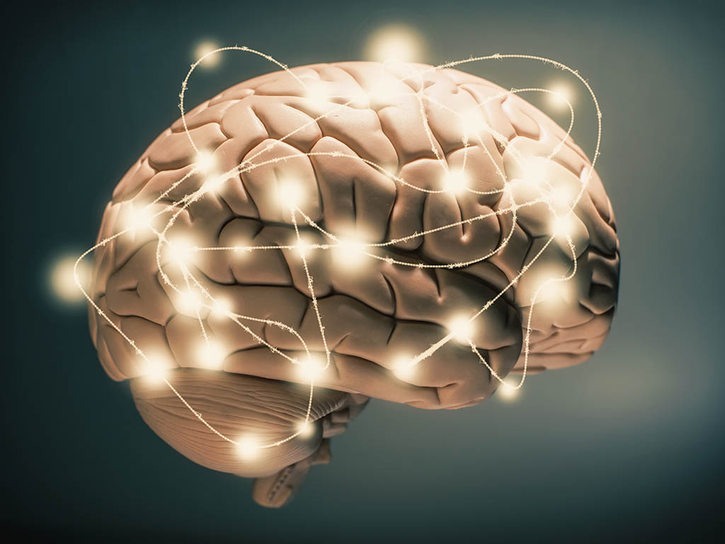
Photo Credit: The Independant Where does human consciousness exist in the brain? Is there a specific location or is consciousness the result of the sum of the entire brain’s activity? Researchers at Vanderbilt examined participants’ brain activity when presented with visual stimuli. Applying Graph Theory, the researchers concluded that consciousness is the result of global activity in the brain rather than focal activity. The study was discussed in a recent article in MedicalXpress:
"Identifying the fingerprints of consciousness in humans would be a significant advancement for basic and medical research, let alone its philosophical implications on the underpinnings of the human experience," said René Marois, professor and chair of psychology at Vanderbilt University and senior author of the study. "Many of the cognitive deficits observed in various neurological diseases may ultimately stem from changes in how information is communicated throughout the brain."
Using graph theory, a branch of mathematics concerned with explaining the interactive links between members of a complex network, such as social networks or flight routes, the researchers aimed to characterize how connections between the various parts of the brain were related to awareness.
"With graph theory, one can ask questions about how efficiently the transportation networks in the United States and Europe are connected via transportation hubs like LaGuardia Airport in New York," Douglass Godwin, graduate student and lead author on the research, said. "We can ask those same questions about brain networks and hubs of neural communication."
Modern theories of the neural basis of consciousness fall generally into two camps: focal and global. Focal theories contend there are specific areas of the brain that are critical for generating consciousness, while global theories argue consciousness arises from large-scale brain changes in activity. This study applied graph theory analysis to adjudicate between these theories.
The researchers recruited 24 members of the university community to participate in a functional magnetic resonance imaging (fMRI) experiment. While in the fMRI scanner, participants were asked to detect a disk that was briefly flashed on a screen. In each trial, participants responded whether they were able to detect the target disk and how much confidence they had in their answer. Experimenters then compared the results of the high-confidence trials during which the target was detected to the trials when it was missed by participants. These were treated as "aware" and "unaware" trials, respectively.
Comparison of aware and unaware trials using conventional fMRI analyses that assess the amplitude of brain activity showed a pattern of results typical of similar studies, with only a few areas of the brain showing more activity during detection of the target than when participants missed seeing it. The present study, however, was interested not simply in what regions might be more activated with awareness, but how they communicate with one another.
Unlike the focal results seen using more conventional analysis methods, the results via this network approach pointed toward a different conclusion. No one area or network of areas of the brain stood out as particularly more connected during awareness of the target; the whole brain appeared to become functionally more connected following reports of awareness.
"We know there are numerous brain networks that control distinct cognitive functions such as attention, language and control, with each node of a network densely interconnected with other nodes of the same network, but not with other networks," Marois said. "Consciousness appears to break down the modularity of these networks, as we observed a broad increase in functional connectivity between these networks with awareness."
The research suggests that consciousness is likely a product of this widespread communication, and that we can only report things that we have seen once they are being represented in the brain in this manner. Thus, no one part of the brain is truly the "seat of the soul," as René Descartes once wrote in a hypothesis about the pineal gland, but rather, consciousness appears to be an emergent property of how information that needs to be acted upon gets propagated throughout the brain.
"We take for granted how unified our experience of the world is. We don't experience separate visual and auditory worlds, it's all integrated into a single conscious experience," Godwin said. "This widespread cross-network communication makes sense as a mechanism by which consciousness gets integrated into that singular world."
Read the full article Here
- Comments (0)
- Memories Fade Without Reinforcement
- By Jason von Stietz
- March 24, 2015
-
Photo Credit: iStockPhotos What happens to our memories? Do they fade over time or do newer ones over shadow them? Researchers from Birmingham and Cambridge, England conducted a study examining how brain patterns found using MRI scans change during a learning task. The researchers concluded that the formation of new memories “actively degraded” older memories. The study was discussed in a recent article in The New York Times:
If old memories stay strong and are merely papered over by new ones, they may be easier to recover. That could be positive for someone trying to remember an acquaintance’s name, but difficult for someone trying to lessen memories of abuse. It could suggest different strategies for easing traumatic memories, evaluating witness testimony about crimes, or helping students study for tests.
Now, a study claims to provide evidence of memory’s weakening by showing that people’s ability to remember something and the pattern of brain activity that thing generates both appear to diminish when a competing memory gets stronger.
Demonstrating sophisticated use of brain scans in memory research, authors of the study, published Monday in the journal Nature Neuroscience, appear to have identified neural fingerprints of specific memories, distinguishing brain activity patterns produced when viewing a picture of a necklace, say, from a picture of binoculars or other objects.
The experiment, conducted by scientists in Birmingham and Cambridge, England, involved several stages with 24 participants first trained to associate words to two unrelated black and white pictures from lists of famous people, ordinary objects or scenes.
They then completed several tasks in a brain scanner. First, they were shown a cue word and asked to recall the image they had been trained to link to that word so that image would become the dominant memory. (For consistency, they were asked to recall the first image they were trained on.)
For example, if the word “sand” was associated first with Marilyn Monroe and then with a hat, scientists wanted participants to indicate that they were recalling Monroe by pressing a button. Each cue word was sprinkled into the test four times, so scientists could see if participants looking at the word “sand” increasingly chose Monroe over the competing memory of the hat. They did.
Next, scientists wanted to see what happened to the hat memory: Did it stay as intact as Monroe although it was not being used, or did it become weaker? To gauge this, scientists showed people two different pictures of Monroe and two hat pictures, asking them which version they had been trained to recognize. If the hat memory had not degraded, scientists reasoned, people would pick the right hat as often as they picked the right Monroe.
To measure success, scientists devised a standard: how well people recalled the correct picture of an unrelated famous person or object. These were images they had been shown early in the study but would have no reason to recall well because they had not been cued to remember them.
For faces, a standard was two Albert Einstein pictures, and people picked the right Monroe about as well as they picked the right Einstein. For objects, a standard was two pictures of goggles. It turned out people were worse at picking the correct hat; they remembered the correct goggles better, even though their memory of goggles had not been reinforced.
Brice Kuhl, a psychology professor at New York University who was not involved in the study, said that strongly suggested that competing memories get weaker, that when people repeatedly pulled out the memory of Monroe in the word test, their recollection of the hat diminished so they did worse at recalling it later.
“You might think it would be better or at least the same” as the standard pictures, he said, “because you’ve just actually had a reminder for the hat, the cue word” in the previous scanner test.
That people had trouble remembering the right hat, he said, makes it less likely the hat memory was simply overshadowed by Monroe. “It’s pretty hard to think that your inability to pick the right hat has anything to do with Marilyn Monroe at that point.”
Next, researchers obtained a “neural signature” of Monroe, the hat and other images by recording brain activity in the prefrontal cortex as participants viewed each picture six times, said a study author, Maria Wimber, a cognitive neuroscientist at the University of Birmingham.
Matching those signatures to brain patterns from the “cue word” test, researchers saw that when the word “sand” was first shown, people’s brains reflected both Monroe and hat patterns, but with subsequent “sand” cues, their brains produced fewer hat traces.
“We watched the memories being suppressed, actively degraded,” said Dr. Wimber, whose collaborators included Michael C. Anderson, a longtime proponent of the memory suppression theory. “It’s not just that the target memories get stronger; the other memories get weaker.”
That interpretation is not necessarily accepted by proponents of memory overshadowing and similar theories.
“I buy that the brain patterns becomes less and less similar to the hat,” said David E. Huber, a professor of psychological and brain sciences at the University of Massachusetts, Amherst, calling the neural signature technique exciting. “Their interpretation of that is that the memory of the hat has been degraded. It’s also possible that increasingly, you’ve learned to think about something other than the hat.”
Dr. Huber noted that while people performed below the standard level at picking the right hat, it was surprising they were not better than standard with Monroe. Dr. Wimber said that had surprised her, too, but her theory is that mental blurring occurs “the more often you bring a picture back to mind,” so details get lost.
Kenneth Norman, a Princeton neuroscientist who was also not involved in the study, said he believed it showed a memory-weakening effect, but that “forgetting is multiply determined, and the two main explanations, they’re not mutually exclusive.”
He said therapeutic applications of memory weakening could include extinguishing fears of something like snakes.
“If you show someone a cartoon image of a snake, cute, funny,” he said, “in the moment you’ve caused liking the snake to overcome not liking the snake. If you want to actually weaken a memory, what you need to do is flush it out. It’s the process of the memory coming to mind as a competitor, but losing the competition.”
Read the full article Here
- Comments (0)
- Eminem: From the Perspective of Neuroscience
- By Jason von Stietz
- March 16, 2015
-

Photo Credit: Getty Images Are the brains of creative geniuses different than the rest of us? Does creativity and schizophrenia really go hand in hand? Recently, neuroscientists gathered to discuss the neuroscience of genius proposed how the brain of someone, such as the popular rapper Eminem, might differ from the typical brain. An article in the Science of Us reviewed the discussion:
Eminem may not have known this when he wrote it, but his line about getting along with the voices inside his head is strangely apt. According to psychologist Scott Barry Kaufman, the brains of creative geniuses and people with schizophrenia are similar in surprising ways. They both have an extremely active precuneus, or area that facilitates daydreaming and free association. The only difference is that unlike people with schizophrenia (and Stan), creative geniuses can distinguish between fantasy and reality.
On Sunday, as part of the 92nd Street Y's Seven Days of Genius series, Kaufman, along with neuroscientist Heather Berlin and science rapper Baba Brinkman, discussed the neuroscience of creativity. During the discussion, they explained a bit about what happens in, say, Eminem's brain when he freestyles versus when he raps from memory.
Here are some highlights:
On genius and consistency:
Kaufman: "It’s a great myth that creative geniuses are always consistently geniuses. A lot of creative geniuses are good marketers and high in narcissism, so it can allow them to appear as though they don't have to work hard at all. But lots of aspects of the creative process resemble what everyday people go through when they’re trying to be creative."On being a screw-up:
Kaufman: "It is important to take risks. Most of Thomas Edison's stuff is really crap. He actually had in his generation some of the worst ideas; he just happened to have one or two things that were the best of all time. So it’s okay to have lots of horrible stuff if you can eventually get it together."On Marshall Mathers:
Berlin: "He probably has more advanced connections in terms of his language areas. Over time, when you practice something, a cognitive skill or a motor skill, you’re developing connections in the brain. So I’m sure his brain would look slightly different."
Kaufman: "I would predict that his dorsolateral prefrontal cortex might be smaller than average. That’s the area of the brain that filters curse words."On rappers' creative states:
Berlin: "The medial prefrontal cortex has increased activation when they're improvising compared to doing a memorized rap, which has to do with the internal generation of new ideas. There's decreased activation in the dorsolateral prefrontal cortex, which has to do with your sense of self. When that part turns down, you lose the filter that makes sure you conform to social norms, which allows for the free flow of information. That can be a really good thing, because on stage consciousness can get in the way."Quotes have been edited for length.
Read the original article Here
- Comments (0)
- Possible Early Indication of Alzheimer's Found
- By Jason von Stietz
- March 6, 2015
-
Photo Credit: Getty Images The earliest signs of Alzheimer’s disease may have been found in a recent study. Researchers from Northwestern University examined the brains of those with Alzheimer’s and the brains of healthy participants. Findings suggested that amyloid protein, which are indicative of the disease, were found in healthy participants in their early 20’s. Cassie Shortsleeve discussed the study and possible protective strategies in a recent article in Yahoo Health:
Alzheimer’s disease may not be just for grandparents to worry about: Groundbreaking new research from Northwestern University has found that amyloid protein — a hallmark of the devastating disease — starts accumulating in brain neurons of people as young as 20 years old.
Scientists believe this is the first time that such changes have been noted in human brains so young. In the study, lead researcher Changiz Geula and his team from the Cognitive Neurology and Alzheimer’s Disease Center at Northwestern University’s Feinberg School of Medicine and team analyzed neurons from the brains of 13 “normal” young people ages 20 to 66; 16 people ages 70 to 99 without dementia; and 21 people with Alzheimer’s, ages 60 to 95.
“It was the age that really that surprised us,” Geula tells Yahoo! Health. “In the young adults, we already see accumulation of amyloids.” This becomes worrisome when amyloid clumps grow in size and abundance. The difference between old and young in this study: While the amyloids themselves were present in younger people — and clumps were present as well — there was more clumping in the aging and Alzheimer’s population, says Geula.
“What this means is these neurons are susceptible to accumulate at a young age, but that the clumping really occurs in aging. During life, the substance needed to make clumps is available. And if you have susceptibility to form clumps, this could worsen.”
So what does this mean for you? First, know this: “In this study, we didn’t have a huge number of brains,” says Geula. “And this doesn’t mean that because young people have a measure of amyloids that everyone is going to get Alzheimer’s. It’s not an alarm.”
But there are susceptibility factors (that science knows a lot about) — and protective factors (that science doesn’t know as much about) — when it comes to Alzheimer’s, Geula says. “We have known for a while that if we want effective therapy for Alzheimer’s, we have to start early. What these findings suggest is the earlier the better.”
So start today and protect yourself — and your brain —with these techniques, no matter your age.
1. Kick bad habits—stat. “We know that general cognitive aging and Alzheimer’s aging can be enhanced by many things — most importantly, general health,” says Geula. In fact, many health factors — diabetes, heart disease, pulmonary function, and obesity — can increase your risk of the disease.
2. Clean up your diet. Healthy habits like eating a Mediterranean diet (rich in nuts, greens, whole grains, fruits and veggies, poultry, and olive oil) have been linked to better cognitive function and a lower risk of Alzheimer’s.
3. Work it out. Exercise has been shown to enhance cognitive abilities in everyone, including the old and even the Alzheimer’s population. “Some animal studies show that the total amount of amyloid can be reduced in animals through enhanced exercise and enriched environment,” says Geula. And not only can exercise keep Alzheimer’s and dementia at bay, some research suggests it can even turn it around. When people with mild cognitive impairment walked on a treadmill for 30 minutes a day, four days a week for 12 weeks, people improved their neural efficiency using fewer mental resources to perform the same task.
4. Better your brain. Mental exercise — keeping your mind engaged and challenged through a variety of exercises — can also help. Research suggests that people who play cards, do crossword puzzles, and challenge themselves mentally on a regular basis are at a lower risk for developing the disease than those who don’t. And thinking on the bright side actually does matter: Negative thoughts can hinder your brain’s ability to think straight and form memories, according to research from King’s College in London.Read the original article Here
- Comments (0)
 Subscribe to our Feed via RSS