Blog
- Schizophrenia Might Be Tied to Humans' Higher Cognitive Abilities Compared to Non-Human Animals
- By Jason von Stietz
- June 30, 2015
-

Photo Credit: Getty Images Research has suggested that non-human animals can suffer from mental illness. For example, dog owners might be correct in believing that their pet suffers from separation anxiety. Furthermore, dolphins and whales have been known to engage in self-mutilating behavior when in captivity. However, humans are the only animals to suffer from schizophrenia. Recent findings have suggested that schizophrenia could be tied to the higher cognitive abilities possessed by humans. A recent article in Scientific American reviewed the research:
Plenty of us have known a dog on Prozac. We have also witnessed the eye rolls that come with the mention of canine psychiatry. Doting pet owners—myself included—ascribe all kinds of questionable psychological ills to our pawed companions. But in fact, the science suggests that numerous nonhuman species do suffer from psychiatric symptoms. Birds obsess; horses on occasion get pathologically compulsive; dolphins and whales, especially those in captivity, self-mutilate. And that thing when your dog woefully watches you pull out of the driveway from the window—that might be DSM-certified separation anxiety. “Every animal with a mind has the capacity to lose hold of it from time to time,” wrote science historian and author Laurel Braitman in her 2014 book Animal Madness.
But at least one mental malady, while common in humans, seems to have spared other animals: schizophrenia, which affects an estimated 0.4 to 1 percent of adults. Although animal models of psychosis exist in laboratories, and odd behavior has been observed in creatures confined to cages, most experts agree that psychosis has not typically been seen in other species, whereas depression, obsessive-compulsive disorder and anxiety traits have been reported in many nonhuman species.
This raises the question of why such a potentially devastating, often lethal disease is still hanging around plaguing humanity. We know from an abundance of recent research that schizophrenia is heavily genetic in origin. One would think that natural selection would have eliminated the genes that predispose to psychosis. A study published earlier this year in Molecular Biology and Evolution provides clues as to how the potential for schizophrenia may have arisen in the human brain and, in doing so, suggests possible treatment targets. It turns out that psychosis may be an unfortunate cost of having a big brain that is capable of complex cognition.
Hotspots in the Human Genome
The study, led by Joel Dudley, a genomics professor at the Icahn School of Medicine at Mount Sinai, proposes that because schizophrenia is relatively prevalent in humans, it perhaps has a complex evolutionary backstory that would explain its persistence and apparent exclusivity to humans. Specifically, Dudley and his colleagues were curious about segments of our genome called human accelerated regions, or HARs, first identified in 2006. HARs are short stretches of DNA that were conserved in other species but underwent rapid evolution in humans following our split with chimpanzees, presumably because they provided some benefit specific to our species. Rather than encoding for proteins themselves, HARs often help to regulate neighboring genes. Because both schizophrenia and HARs appear to be, for the most part, human-specific, the researchers wondered if there might be a connection between the two.To find out, Dudley and his colleagues used data culled from the Psychiatric Genomics Consortium, a massive study identifying genetic variants associated with schizophrenia. They first assessed whether schizophrenia-related genes sit close to HARs along the human genome—closer than would be expected by chance. It turns out they do, suggesting that HARs play a role in regulating genes contributing to schizophrenia. Furthermore, by comparing the patterns of change in humans and chimpanzees, it was revealed that HAR-associated schizophrenia genes were under stronger evolutionary selective pressure than other schizophrenia genes. This observation implies that the human variants of these genes are essential to us in some way, despite the risk they harbor.
To help understand what these benefits might be, Dudley's group then turned to gene expression profiles. Gene sequencing provides an organism's genome sequence, but gene expression profiling reveals where and when in the body certain genes are active. Dudley's team found that HAR-associated schizophrenia genes are found in regions of the genome that influence other genes expressed in the prefrontal cortex, a brain region just behind the forehead that is involved in higher-order thinking. Impaired function in the prefrontal cortex is thought to contribute to psychosis.
They also found that these culprit genes are involved in various key human neurological functions within the prefrontal cortex, including the transmission of the neurotransmitter GABA across a synapse from one neuron to another. GABA serves as an inhibitor or regulator of neuronal activity, in part by suppressing dopamine in certain parts of the brain. In schizophrenia, GABA appears to malfunction, and dopamine runs wild, contributing to the hallucinations, delusions and disorganized thinking that are common to psychosis. In other words, the schizophrenic brain lacks restraint.
“The ultimate goal of the study was to see if evolution may help provide additional insights into the genetic architecture of schizophrenia so that we can better understand and diagnose the disease,” Dudley explains. Identifying which genes are most implicated in schizophrenia and how they are expressed could lead to more effective therapies such as those influencing the function of GABA.
When Bigger Isn't Better
Dudley's findings offer a possible explanation for why schizophrenia arose in humans in the first place and why it does not seem to occur in other animals. “It's been suggested,” Dudley explains, “that the emergence of human speech and language bears a relationship with schizophrenia genetics and, incidentally, autism.” Indeed, language dysfunction—typified by disorganized speech or jumping from one topic to another—is a feature of schizophrenia, and GABA is critical to speech, language and many other aspects of higher-order cognition. “The fact that our evolutionary analysis converged on GABA function in the prefrontal cortex seems to tell an evolutionary story connecting schizophrenia risk with intelligence.”Put another way, with complicated, highly social human thought—and the complicated genetics at the root of higher cognition—perhaps there is just more that can go wrong: complex function begets complex malfunction.
Dudley is careful not to exaggerate the evolutionary implications of his work. “It is important to note that our study was not specifically designed to evaluate an evolutionary trade-off,” he observes, “but our findings support the hypothesis that evolution of our advanced cognitive abilities may have come at a cost—a predisposition to schizophrenia.” He also acknowledges that the new work did not identify any “smoking gun genes” and that schizophrenia genetics is profoundly complex. Still, Dudley feels that evolutionary genetic analysis can help identify the most relevant genes and pathological mechanisms at play in schizophrenia and possibly other mental illnesses that preferentially affect humans—that is, neurodevelopmental disorders related to higher cognition and GABA activity, including autism and attention-deficit/hyperactivity disorder.
In fact, a study published online this past March in Molecular Psychiatry reported a link between gene variants associated with autism spectrum disorder and better cognitive function in the general population—specifically, enhanced general cognitive ability, memory and verbal intelligence. “It would suggest that some of these variants can have beneficial effects on cognition,” says lead author Toni-Kim Clarke of the University of Edinburgh. The findings might also help explain why individuals with autism sometimes exhibit unusual cognitive gifts.
Clarke's findings support Dudley's speculation that higher cognition might have come at a price. As we broke away from our primate cousins, our genomes—HARs especially—hastily evolved, granting us an increasing cache of abilities that other species lack. In doing so, they may have left our brains prone to occasional complex dysfunction—but also capable of biomedical research aimed at one day curing the ailing brain.
Read original article Here
- Comments (1)
- Cortical and Subcortical Synchrony Involved in Hypersensitivity to Sensory Stimuli in ASD
- By Jason von Stietz
- June 23, 2015
-

Photo Credit: Getty Images A well known symptom of autism spectrum disorder (ASD) is hyper sensitivity to sensory stimuli. Researchers at the Netherlands Institute for Neuroscience examined fMRI data from the largest neuroimaging database on ASD. Findings indicated that highly synchronized activity between cortical and subcortical brain activity is related to hypersensitivity to sensory stimuli in people with ASD. A recent article in NeuroScientistNews discusses the study:
The increased interaction between cortical and subcortical brain regions highlights the central role of hypersensitivity and other sensory symptoms in defining autism spectrum disorder (ASD). This is presented in research performed by a team led by Christian Keysers and Leonardo Cerliani at the Netherlands Institute for Neuroscience in Amsterdam. This finding provides a key to understand the often underestimated sensory hypersensitivity in autism and to seed a scientific understanding of how to tackle this hypersensitivity. The research is published in JAMA Psychiatry.
People with ASD are known for their unusual behavior in the social environment. Moreover, they often report other traits, linked to the sensory environment: for instance the ability to perceive small details in a picture or to detect a very soft sound coming from a distance. "This hypersensitivity, however, is not always a gift: being captured by the myriad of sensory stimuli we continuously receive from the environment can be distracting and even overwhelming, and prevents us to focus on what we care most," says Cerliani. The scientists present in their research that increased interaction between the cortical ad subcortical brain regions is at the root of this hypersensitivity.
Brain activity
Brain regions that are strongly coupled have brain activity that goes up and down together, even while relaxing, while regions that are not coupled will have their brain activity fluctuate independently from each other. By comparing how this spontaneous brain activity synchronizes across various brain regions the team identified an abnormally high synchrony between the sensory cortices involved in perception and subcortical regions relaying information from the sensory organs to the cortex. They found that a higher synchrony was associated with a higher severity of autistic traits.
"During the development from childhood to adolescence, the spontaneous activity of cortical regions involved in basic sensory perception decouples from the activity of subcortical structures relaying sensory information from the sensory organs to the cortex," explains Keysers. "This decoupling is thought to reflect the increasing ability to block out irrelevant sensory information from perception, allowing people to focus on their stream of thoughts and actions. In ASD this process appears to be altered: their sensory cortex appears to be abnormally coupled to subcortical structures."
Large database
The team made these observations using resting-state functional magnetic resonance imaging (fMRI) data from the largest neuroimaging database on ASD aggregated so far: the ABIDE, founded and coordinated by Dr. Adriana Di Martino, Dr. Stuart Mostofsky and Dr. Michael Milham. The team of Keysers and Cerliani also contributed to the aggregation of this database with the neuroimaging data acquired by Dr. Marc Thioux.
Autism Spectrum Disorder
ASD is an umbrella name for a number of developmental disorders, including classic autism and Asperger syndrome. The number of people with ASD is almost eightfold in the last twenty years and is seen in more than 1% of the children.
Read the original article Here
- Comments (0)
- Sleep and Impulse Control Issues Related to Spindling Excessive Beta
- By Jason von Stietz
- June 12, 2015
-
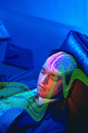
Photo Credit: Getty Images Can EEG phenotypes predict issues with sleep and with impulse control? Brain Science International’s Jay Gunkelman QEEG-D recently co-authored an article on the relationship between the EEG-beta phenotype and behavior. The findings suggested that frontal spindling beta related to sleep maintenance issues and impulse control problems. The article was published in the peer reviewed journal Neuropsychiatric Electrophysiology:
Abstract
Background
In 2009 the United States National Institute of Mental Health (NIMH) introduced the Research Domain Criteria (RDoC) project, which intends to explicate fundamental bio-behavioral dimensions that cut across heterogeneous disorder categories in psychiatry. One major research domain is defined by arousal and regulatory systems.
Methods
In this study we aimed to investigate the relation between arousal systems (EEG-beta phenotypes also referred to as spindling excessive beta (SEB), beta spindles or sub-vigil beta) and the behavioral dimensions: insomnia, impulsivity/hyperactivity and attention. This analysis is conducted within a large and heterogeneous outpatient psychiatric population, in order to verify if EEG-beta phenotypes are an objective neurophysiological marker for psychopathological properties shared across psychiatric disorders.
Results
SEBs had an occurrence between 0–10.8% with a maximum occurrence at frontal and central locations, with similar topography for the heterogeneous sample as well as a more homogenous ADHD subgroup. Patients with frontal SEBs only, had significantly higher impulsivity/hyperactivity (specifically on impulse control items) and insomnia complaints with medium effect sizes.
Conclusions
Item level and mediation analysis revealed that sleep maintenance problems explained both frontal SEB EEG patterns (in line with SEB as a sub-vigil or hypoarousal EEG pattern) as well as the impulse control problems. These data thus suggest that frontal SEB might be regarded as a state marker caused by sleep maintenance problems, with concurrent impulse control problems. However, future longitudinal studies should investigate this state-trait issue further and replicate these findings Also studies manipulating SEB by for example neurofeedback and measuring consequent changes in sleep and impulse control could shed further light on this issue.
Read the article online Here
- Comments (0)
- Transcranial Direct Current Stimulation Improves Memory in People with Schizophrenia
- By Jason von Stietz
- June 5, 2015
-
Photo Credit: Getty Images Researchers at Johns Hopkins University found that stimulating the brain with a weak electrical current can lead to improvements in the short-term memory of people with schizophrenia. According to the researchers, transcranial direct current stimulation led to significant improvements in visual and verbal memory. The study was discussed in a recent article in NeuroScientistNews:
Lightly stimulating the brain with electricity may improve short-term memory in people with schizophrenia, according to a new study by researchers at the Johns Hopkins University School of Medicine.
The procedure, known as transcranial direct current stimulation, involves placing sponge-covered electrodes on the head and passing a weak electrical current between them. It is widely regarded as safe, and the procedure is being studied as a treatment for depression and Alzheimer's-related memory loss, and to enhance recovery following strokes. David Schretlen, Ph.D., a professor of psychiatry and behavioral sciences at the Johns Hopkins University School of Medicine, reasoned that this type of brain stimulation might ease some of the cognitive difficulties that afflict people with schizophrenia.
"Cognitive impairment is as ubiquitous as hallucinations in schizophrenia, yet medications only treat the hallucinations. So even with medication, affected individuals often remain very disabled," Schretlen says. His hope is that transcranial direct current stimulation could give people with schizophrenia a shot at leading a more normal life.
To test that possibility, Schretlen and five Johns Hopkins colleagues targeted the left dorsolateral prefrontal cortex, which plays an important role in short-term or working memory and is abnormal in people with schizophrenia. Interestingly, parents, siblings and children of people with schizophrenia show some of the same abnormalities to a lesser degree.
Schretlen recruited 11 participants: five adults with confirmed schizophrenia and six of their close relatives. Each participant received two 30-minute treatments -- one using a negative electrical charge, which the researchers thought might prove beneficial -- and the other using a positive charge as a control. During and after each treatment, participants completed a battery of cognitive tests. On tests of verbal and visual working memory, participants performed significantly better after receiving a negative charge, and the effects were "surprisingly strong," says Schretlen.
Schretlen also tested participants' verbal fluency, or word retrieval, during the treatment. People with schizophrenia often struggle to find the right words, Schretlen explains. Because the prefrontal cortex contains a brain region responsible for word retrieval, Schretlen thought transcranial direct current stimulation might help. To test that theory, he gave participants a minute to list things they could buy in a supermarket. Most people taking the test rattle off items in categories, naming fruits, then vegetables, then dairy products, for example. Schretlen found that while participants did not rattle off more words, they did better at the challenging task of switching between categories after a negatively charged treatment.
The stimulation "was associated with better performance on working memory and subtle changes in word retrieval," Schretlen says.
Schretlen is now studying transcranial direct current stimulation in a larger sample of patients using repeated sessions of stimulation, which he hopes will induce lasting benefits.
"What's nice about transcranial direct current stimulation is that it's so benign. There are no bad side effects," Schretlen says. "If it enables people with schizophrenia to think more clearly, it would make a huge contribution to the treatment of this devastating illness."
Read the original aricle Here
- Comments (0)
 Subscribe to our Feed via RSS