Blog
- Brain Circuitry Related to Context Processing Involved in PTSD
- By Jason von Stietz, M.A.
- October 31, 2016
-
Why are some individuals vulnerable to PTSD when others are not? Researchers at University of Michigan theorize that dysregulation in the brain’s circuity related to context processing, hippocampal-prefrontal-thalamic circuitry, is at the core of PTSD. The theory was discussed in a recent article in Medical Xpress:
All experts in the field now agree that PTSD indeed has its roots in very real, physical processes within the brain - and not in some sort of psychological "weakness". But no clear consensus has emerged about what exactly has gone "wrong" in the brain.
In a Perspective article published this week in Neuron, a pair of University of Michigan Medical School professors—who have studied PTSD from many angles for many years—put forth a theory of PTSD that draws from and integrates decades of prior research. They hope to stimulate interest in the theory and invite others in the field to test it.
The bottom line, they say, is that people with PTSD appear to suffer from disrupted context processing. That's a core brain function that allows people and animals to recognize that a particular stimulus may require different responses depending on the context in which it is encountered. It's what allows us to call upon the "right" emotional or physical response to the current encounter.
A simple example, they write, is recognizing that a mountain lion seen in the zoo does not require a fear or "flight" response, while the same lion unexpectedly encountered in the backyard probably does.
For someone with PTSD, a stimulus associated with the trauma they previously experienced - such as a loud noise or a particular smell—triggers a fear response even when the context is very safe. That's why they react even if the noise came from the front door being slammed, or the smell comes from dinner being accidentally burned on the stove.
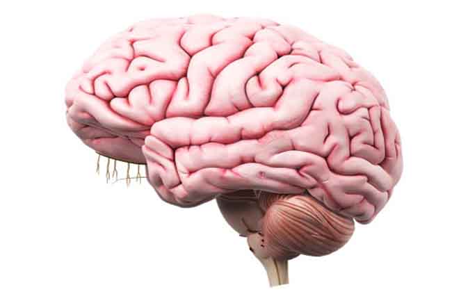
Context processing involves a brain region called the hippocampus, and its connections to two other regions called the prefrontal cortex and the amygdala. Research has shown that activity in these brain areas is disrupted in PTSD patients. The U-M team thinks their theory can unify wide-ranging evidence by showing how a disruption in this circuit can interfere with context processing and can explain most of the symptoms and much of the biology of PTSD.
"We hope to put some order to all the information that's been gathered about PTSD from studies of human patients, and of animal models of the condition," says Israel Liberzon, M.D., a professor of psychiatry at U-M and a researcher at the VA Ann Arbor Healthcare System who also treats veterans with PTSD. "We hope to create a testable hypothesis, which isn't as common in mental health research as it should be. If this hypothesis proves true, maybe we can unravel some of the underlying pathophysiological processes, and offer better treatments."
Liberzon and his colleague, James Abelson, M.D., Ph.D., describe in their piece models of PTSD that have emerged in recent years, and lay out the evidence for each. The problem, they say, is that none of these models sufficiently explains the various symptoms seen in patients, nor all of the complex neurobiological changes seen in PTSD and in animal models of this disorder.
The first model, abnormal fear learning, is rooted in the amygdala - the brain's 'fight or flight' center that focuses on response to threats or safe environments. This model emerged from work on fear conditioning, fear extinction and fear generalization.
The second, exaggerated threat detection, is rooted in the brain regions that figure out what signals from the environment are "salient", or important to take note of and react to. This model focuses on vigilance and disproportionate responses to perceived threats.
The third, involving executive function and regulation of emotions, is mainly rooted in the prefrontal cortex - the brain's center for keeping emotions in check and planning or switching between tasks.
By focusing only on the evidence bolstering one of these theories, researchers may be "searching under the streetlight", says Liberzon. "But if we look at all of it in the light of context processing disruption, we can explain why different teams have seen different things. They're not mutually exclusive."
The main thing, says Liberzon, is that "context is not only information about your surroundings - it's pulling out the correct emotion and memories for the context you are in."
A deficit in context processing would lead PTSD patients to feel "unmoored" from the world around them, unable to shape their responses to fit their current contexts. Instead, their brains would impose an "internalized context"—one that always expects danger—on every situation.
This type of deficit, arising in the brain from a combination of genetics and life experiences, may create vulnerability to PTSD in the first place, they say. After trauma, this would generate symptoms of hypervigilance, sleeplessness, intrusive thoughts and dreams, and inappropriate emotional and physical outbursts.
Liberzon and Abelson think that testing the context processing theory will enhance understanding of PTSD, even if all of its details are not verified. They hope the PTSD community will help them pursue the needed research, in PTSD patients and in animal models. They put forth specific ideas in the Neuron paper to encourage that, and are embarking on such research themselves.
The U-M/VA team is currently recruiting people with PTSD - whether veterans or not - for studies involving brain imaging and other tests.
In the meantime, they note that there is a growing set of therapeutic tools that can help patients with PTSD, such as cognitive behavioral therapy mindfulness training and pharmacological approaches. These may work by helping to anchor PTSD patients in their current environment, and may prove more effective as researchers learn how to specifically strengthen context processing capacities in the brain.
Read the original article here
- Comments (0)
- EEG Can Be Used For Cyber Security
- By Jason von Stietz, M.A.
- October 29, 2016
-
Getty Images Finger print scans are widely used as a method of proving identification and can even be used to access a secured cyber system. However, finger print scans can be stolen or replicated. So, what is the next step in cyber security? Researchers are currently investigating how EEG can be used as a means of biometric authentication. The research was discussed in a recent article in Neuroscience News:
Cyber security and authentication have been under attack in recent months as, seemingly every other day, a new report of hackers gaining access to private or sensitive information comes to light. Just recently, more than 500 million passwords were stolen when Yahoo revealed its security was compromised.
Securing systems has gone beyond simply coming up with a clever password that could prevent nefarious computer experts from hacking into your Facebook account. The more sophisticated the system, or the more critical, private information that system holds, the more advanced the identification system protecting it becomes.
Fingerprint scans and iris identification are just two types of authentication methods, once thought of as science fiction, that are in wide use by the most secure systems. But fingerprints can be stolen and iris scans can be replicated. Nothing has proven foolproof from being subject to computer hackers.
“The principal argument for behavioral, biometric authentication is that standard modes of authentication, like a password, authenticates you once before you access the service,” said Abdul Serwadda a cybersecurity expert and assistant professor in the Department of Computer Science at Texas Tech University.
“Now, once you’ve accessed the service, there is no other way for the system to still know it is you. The system is blind as to who is using the service. So the area of behavioral authentication looks at other user-identifying patterns that can keep the system aware of the person who is using it. Through such patterns, the system can keep track of some confidence metric about who might be using it and immediately prompt for reentry of the password whenever the confidence metric falls below a certain threshold.”
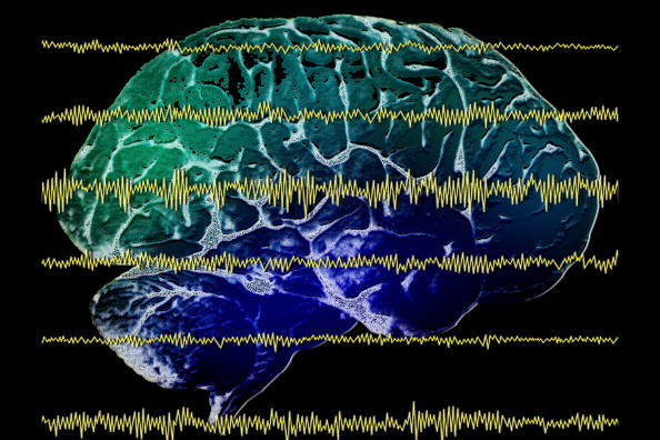
One of those patterns that is growing in popularity within the research community is the use of brain waves obtained from an electroencephalogram, or EEG. Several research groups around the country have recently showcased systems which use EEG to authenticate users with very high accuracy.
However, those brain waves can tell more about a person than just his or her identity. It could reveal medical, behavioral or emotional aspects of a person that, if brought to light, could be embarrassing or damaging to that person. And with EEG devices becoming much more affordable, accurate and portable and applications being designed that allows people to more readily read an EEG scan, the likelihood of that happening is dangerously high.
“The EEG has become a commodity application. For $100 you can buy an EEG device that fits on your head just like a pair of headphones,” Serwadda said. “Now there are apps on the market, brain-sensing apps where you can buy the gadget, download the app on your phone and begin to interact with the app using your brain signals. That led us to think; now we have these brain signals that were traditionally accessed only by doctors being handled by regular people. Now anyone who can write an app can get access to users’ brain signals and try to manipulate them to discover what is going on.”
That’s where Serwadda and graduate student Richard Matovu focused their attention: attempting to see if certain traits could be gleaned from a person’s brain waves. They presented their findings recently to the Institute of Electrical and Electronics Engineers (IEEE) International Conference on Biometrics.
Brain waves and cybersecuritySerwadda said the technology is still evolving in terms of being able to use a person’s brain waves for authentication purposes. But it is a heavily researched field that has drawn the attention of several federal organizations. The National Science Foundation (NSF), funds a three-year project on which Serwadda and others from Syracuse University and the University of Alabama-Birmingham are exploring how several behavioral modalities, including EEG brain patterns, could be leveraged to augment traditional user authentication mechanisms.
“There are no installations yet, but a lot of research is going on to see if EEG patterns could be incorporated into standard behavioral authentication procedures,” Serwadda said
Assuming a system uses EEG as the modality for user authentication, typically for such a system, all variables have been optimized to maximize authentication accuracy. A selection of such variables would include:
The features used to build user templates.
The signal frequency ranges from which features are extracted.
The regions of the brain on which the electrodes are placed, among other variables.Under this assumption of a finely tuned authentication system, Serwadda and his colleagues tackled the following questions:
If a malicious entity were to somehow access templates from this authentication-optimized system, would he or she be able to exploit these templates to infer non-authentication-centric information about the users with high accuracy?
In the event that such inferences are possible, which attributes of template design could reduce or increase the threat?Turns out, they indeed found EEG authentication systems to give away non-authentication-centric information. Using an authentication system from UC-Berkeley and a variant of another from a team at Binghamton University and the University of Buffalo, Serwadda and Matovu tested their hypothesis, using alcoholism as the sensitive private information which an adversary might want to infer from EEG authentication templates.
In a study involving 25 formally diagnosed alcoholics and 25 non-alcoholic subjects, the lowest error rate obtained when identifying alcoholics was 25 percent, meaning a classification accuracy of approximately 75 percent.
When they tweaked the system and changed several variables, they found that the ability to detect alcoholic behavior could be tremendously reduced at the cost of slightly reducing the performance of the EEG authentication system.
Motivation for discovery
Serwadda’s motivation for proving brain waves could be used to reveal potentially harmful personal information wasn’t to improve the methods for obtaining that information. It’s to prevent it.
To illustrate, he gives an analogy using fingerprint identification at an airport. Fingerprint scans read ridges and valleys on the finger to determine a person’s unique identity, and that’s it.
In a hypothetical scenario where such systems could only function accurately if the user’s finger was pricked and some blood drawn from it, this would be problematic because the blood drawn by the prick could be used to infer things other than the user’s identity, such as whether a person suffers from certain diseases, such as diabetes.
Given the amount of extra information that EEG authentication systems are able glean about the user, current EEG systems could be likened to the hypothetical fingerprint reader that pricks the user’s finger. Serwadda wants to drive research that develops EEG authentication systems that perform the intended purpose while revealing minimal information about traits other than the user’s identity in authentication terms.
Currently, in the vast majority of studies on the EEG authentication problem, researchers primarily seek to outdo each other in terms of the system error rates. They work with the central objective of designing a system having error rates which are much lower than the state-of-the-art. Whenever a research group develops or publishes an EEG authentication system that attains the lowest error rates, such a system is immediately installed as the reference point.
A critical question that has not seen much attention up to this point is how certain design attributes of these systems, in other words the kinds of features used to formulate the user template, might relate to their potential to leak sensitive personal information. If, for example, a system with the lowest authentication error rates comes with the added baggage of leaking a significantly higher amount of private information, then such a system might, in practice, not be as useful as its low error rates suggest. Users would only accept, and get the full utility of the system, if the potential privacy breaches associated with the system are well understood and appropriate mitigations undertaken.
But, Serwadda said, while the EEG is still being studied, the next wave of invention is already beginning.
“In light of the privacy challenges seen with the EEG, it is noteworthy that the next wave of technology after the EEG is already being developed,” Serwadda said. “One of those technologies is functional near-infrared spectroscopy (fNIRS), which has a much higher signal-to-noise ratio than an EEG. It gives a more accurate picture of brain activity given its ability to focus on a particular region of the brain.”
The good news, for now, is fNIRS technology is still quite expensive; however there is every likelihood that the prices will drop over time, potentially leading to a civilian application to this technology. Thanks to the efforts of researchers like Serwadda, minimizing the leakage of sensitive personal information through these technologies is beginning to gain attention in the research community.
“The basic idea behind this research is to motivate a direction of research which selects design parameters in such a way that we not only care about recognizing users very accurately but also care about minimizing the amount of sensitive personal information it can read,” Serwadda said.
Read the original article here
- Comments (0)
- Brain Computer Interface Generates Touch Sensation For Paralyzed Man
- By Jason von Stietz, M.A.
- October 21, 2016
-
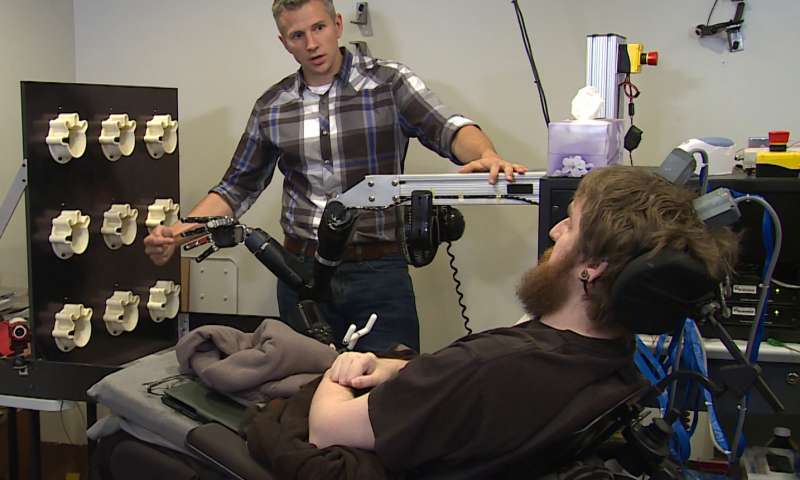
Credit: UPMC/Pitt Healt Sciences Media Relations The sensation of touch is a critical aspect in everyday functioning. However, until recently prosthetic limbs have been unable to recreate the sense of touch for their users. Researcher at the University of Pittsburgh recently examined the use of microelectrodes implanted into the somatosensory cortex of a person with a spinal cord injury, which generate sensations of touch perceived as coming from the person’s own paralyzed limb. The study was discussed in a recent article in Medical Xpress:
Imagine being in an accident that leaves you unable to feel any sensation in your arms and fingers. Now imagine regaining that sensation, a decade later, through a mind-controlled robotic arm that is directly connected to your brain.
That is what 28-year-old Nathan Copeland experienced after he came out of brain surgery and was connected to the Brain Computer Interface (BCI), developed by researchers at the University of Pittsburgh and UPMC. In a study published online today in Science Translational Medicine, a team of experts led by Robert Gaunt, Ph.D., assistant professor of physical medicine and rehabilitation at Pitt, demonstrated for the first time ever in humans a technology that allows Mr. Copeland to experience the sensation of touch through a robotic arm that he controls with his brain.
"The most important result in this study is that microstimulation of sensory cortex can elicit natural sensation instead of tingling," said study co-author Andrew B. Schwartz, Ph.D., distinguished professor of neurobiology and chair in systems neuroscience, Pitt School of Medicine, and a member of the University of Pittsburgh Brain Institute. "This stimulation is safe, and the evoked sensations are stable over months. There is still a lot of research that needs to be carried out to better understand the stimulation patterns needed to help patients make better movements."
This is not the Pitt-UPMC team's first attempt at a BCI. Four years ago, study co-author Jennifer Collinger, Ph.D., assistant professor, Pitt's Department of Physical Medicine and Rehabilitation, and research scientist for the VA Pittsburgh Healthcare System, and the team demonstrated a BCI that helped Jan Scheuermann, who has quadriplegia caused by a degenerative disease. The video of Scheuermann feeding herself chocolate using the mind-controlled robotic arm was seen around the world. Before that, Tim Hemmes, paralyzed in a motorcycle accident, reached out to touch hands with his girlfriend.
But the way our arms naturally move and interact with the environment around us is due to more than just thinking and moving the right muscles. We are able to differentiate between a piece of cake and a soda can through touch, picking up the cake more gently than the can. The constant feedback we receive from the sense of touch is of paramount importance as it tells the brain where to move and by how much.
For Dr. Gaunt and the rest of the research team, that was the next step for the BCI. As they were looking for the right candidate, they developed and refined their system such that inputs from the robotic arm are transmitted through a microelectrode array implanted in the brain where the neurons that control hand movement and touch are located. The microelectrode array and its control system, which were developed by Blackrock Microsystems, along with the robotic arm, which was built by Johns Hopkins University's Applied Physics Lab, formed all the pieces of the puzzle.
In the winter of 2004, Mr. Copeland, who lives in western Pennsylvania, was driving at night in rainy weather when he was in a car accident that snapped his neck and injured his spinal cord, leaving him with quadriplegia from the upper chest down, unable to feel or move his lower arms and legs, and needing assistance with all his daily activities. He was 18 and in his freshman year of college pursuing a degree in nanofabrication, following a high school spent in advanced science courses.
He tried to continue his studies, but health problems forced him to put his degree on hold. He kept busy by going to concerts and volunteering for the Pittsburgh Japanese Culture Society, a nonprofit that holds conventions around the Japanese cartoon art of anime, something Mr. Copeland became interested in after his accident.
Right after the accident he had enrolled himself on Pitt's registry of patients willing to participate in clinical trials. Nearly a decade later, the Pitt research team asked if he was interested in participating in the experimental study.
After he passed the screening tests, Nathan was wheeled into the operating room last spring. Study co-investigator and UPMC neurosurgeon Elizabeth Tyler-Kabara, M.D., Ph.D., assistant professor, Department of Neurological Surgery, Pitt School of Medicine, implanted four tiny microelectrode arrays each about half the size of a shirt button in Nathan's brain. Prior to the surgery, imaging techniques were used to identify the exact regions in Mr. Copeland's brain corresponding to feelings in each of his fingers and his palm.
"I can feel just about every finger—it's a really weird sensation," Mr. Copeland said about a month after surgery. "Sometimes it feels electrical and sometimes its pressure, but for the most part, I can tell most of the fingers with definite precision. It feels like my fingers are getting touched or pushed."
At this time, Mr. Copeland can feel pressure and distinguish its intensity to some extent, though he cannot identify whether a substance is hot or cold, explains Dr. Tyler-Kabara.
Michael Boninger, M.D., professor of physical medicine and rehabilitation at Pitt, and senior medical director of post-acute care for the Health Services Division of UPMC, recounted how the Pitt team has achieved milestone after milestone, from a basic understanding of how the brain processes sensory and motor signals to applying it in patients
"Slowly but surely, we have been moving this research forward. Four years ago we demonstrated control of movement. Now Dr. Gaunt and his team took what we learned in our tests with Tim and Jan—for whom we have deep gratitude—and showed us how to make the robotic arm allow its user to feel through Nathan's dedicated work," said Dr. Boninger, also a co-author on the research paper.
Dr. Gaunt explained that everything about the work is meant to make use of the brain's natural, existing abilities to give people back what was lost but not forgotten.
"The ultimate goal is to create a system which moves and feels just like a natural arm would," says Dr. Gaunt. "We have a long way to go to get there, but this is a great start."
Read the original article Here
- Comments (0)
 Subscribe to our Feed via RSS