Blog
- Can Fast fMRI Monitor Brain Activity of Thoughts?
- By Jason von Stietz, M.A.
- March 30, 2017
-
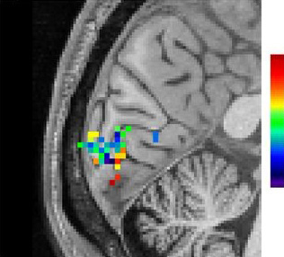
Photo Credit: Lewis et al Can fMRI detect human thought? Researchers from Harvard utilized recent developments in fast fMRI techniques to examine rapid oscillations of brain activity during human thought. Researchers showed participants rapidly oscillating images and monitored the rapid oscillations of brain activity in the visual cortex. These findings ark the first steps towards better studying the neural networks as they produce human thought. The study was discussed funded by the National Institutes for Health and discussed in a recent press release:
By significantly increasing the speed of functional MRI (fMRI), NIBIB-funded researchers have been able to image rapidly fluctuating brain activity during human thought. fMRI measures changes in blood oxygenation, which were previously thought to be too slow to detect the subtle neuronal activity associated with higher order brain functions. The new discovery that fast fMRI can detect rapid brain oscillations is a significant step towards realizing a central goal of neuroscience research: mapping the brain networks responsible for human cognitive functions such as perception, attention, and awareness.
“A critical aim of the President’s BRAIN Initiative1 is to move neuroscience into a new realm where we can identify and track functioning neural networks non-invasively,” explains Guoying Liu, Ph.D., Director of the NIBIB program in Magnetic Resonance Imaging. “This work demonstrates the potential of fMRI for mapping healthy neural networks as well as those that may contribute to neurological diseases such as dementia and other mental health disorders, which are significant national and global health problems.”
fMRI works by detecting local increases in oxygen as blood is delivered to a working part of the brain. The technique has been instrumental for identifying which areas in the brain control functions such as vision, hearing, or touch. However, standard fMRI can only detect the blood flow coming to replenish an area of the brain several seconds after it has performed a function. It was generally accepted that this was the limit of what could be detected by fMRI—identification of a region in the brain that had responded to a large stimulus, such as a continuous 30 second “blast” of bright light.
Combining several new techniques, Jonathan R. Polimeni, Ph.D., senior author of the study, and his colleagues at Harvard’s Athinoula A. Martinos Center for Biomedical Imaging, applied fast fMRI in an effort to track neuronal networks that control human thought processes, and found that they could now measure rapidly oscillating brain activity. The results of this groundbreaking work are reported in the October 2016 issue of the Proceedings of the National Academy of Sciences.2
The researchers used fast fMRI in human volunteers observing a rapidly fluctuating checkerboard pattern. The fast fMRI was able to detect the subtle and very rapid oscillations in cerebral blood flow in the brain’s visual cortex as the volunteers observed the changing pattern.
“The oscillating checkerboard pattern is a more “naturalistic” stimulus, in that its timing is similar to the very subtle neural oscillations made during normal thought processes,” explains Polimeni. “The fast fMRI detects the induced neural oscillations that allow the brain to understand what the eye is observing --- the changing checkerboard pattern. These subtle oscillations were completely undetectable with standard fMRI. This exciting result opens the possibility of using fast fMRI to image neural networks as they guide the process of human thought.”
One such possibility is suggested by first author of the study Laura D. Lewis, Ph.D. “This technique now gives us a method for obtaining much more detailed information about the complex brain activity that takes place during sleep, as well as other dynamic switches in brain states, such as when under anesthesia and during hallucinations.”
Concludes Polimeni, “It had always been thought that fMRI had the potential to play a major role in these types of studies. Meaningful progress in cognitive neuroscience depends on mapping patterns of brain activity, which are constantly and rapidly changing with every experience we have. Thus, we are extremely excited to see our work contribute significantly to achieving this goal.”
Read the original article Here
- Comments (2)
- Interhemispheric Connectivity Related to Creativity
- By Jason von Stietz, M.A.
- March 23, 2017
-
Shutterstock Popular media often suggests that the “right brain” is more associated with creativity and artistic ability. Researchers at Duke University and University of Padova examined the relationship diffusor tensor imaging brain scans and measures of creativity in 68 participants. Findings indicated that greater interhemispheric connectivity related to higher creativity. The study was discussed in a recent article in Medical Xpress:
For the study, statisticians David Dunson of Duke University and Daniele Durante of the University of Padova analyzed the network of white matter connections among 68 separate brain regions in healthy college-age volunteers.
The brain's white matter lies underneath the outer grey matter. It is composed of bundles of wires, or axons, which connect billions of neurons and carry electrical signals between them.
A team led by neuroscientist Rex Jung of the University of New Mexico collected the data using an MRI technique called diffusion tensor imaging, which allows researchers to peer through the skull of a living person and trace the paths of all the axons by following the movement of water along them. Computers then comb through each of the 1-gigabyte scans and convert them to three-dimensional maps—wiring diagrams of the brain.
Jung's team used a combination of tests to assess creativity. Some were measures of a type of problem-solving called "divergent thinking," or the ability to come up with many answers to a question. They asked people to draw as many geometric designs as they could in five minutes. They also asked people to list as many new uses as they could for everyday objects, such as a brick or a paper clip. The participants also filled out a questionnaire about their achievements in ten areas, including the visual arts, music, creative writing, dance, cooking and science.
The responses were used to calculate a composite creativity score for each person.
Dunson and Durante trained computers to sift through the data and identify differences in brain structure.
They found no statistical differences in connectivity within hemispheres, or between men and women. But when they compared people who scored in the top 15 percent on the creativity tests with those in the bottom 15 percent, high-scoring people had significantly more connections between the right and left hemispheres.
The differences were mainly in the brain's frontal lobe.
Dunson said their approach could also be used to predict the probability that a person will be highly creative simply based on his or her brain network structure. "Maybe by scanning a person's brain we could tell what they're likely to be good at," Dunson said.
The study is part of a decade-old field, connectomics, which uses network science to understand the brain. Instead of focusing on specific brain regions in isolation, connectomics researchers use advanced brain imaging techniques to identify and map the rich, dense web of links between them.
Dunson and colleagues are now developing statistical methods to find out whether brain connectivity varies with I.Q., whose relationship to creativity is a subject of ongoing debate.
In collaboration with neurology professor Paul Thompson at the University of Southern California, they're also using their methods for early detection of Alzheimer's disease, to help distinguish it from normal aging.
By studying the patterns of interconnections in healthy and diseased brains, they and other researchers also hope to better understand dementia, epilepsy, schizophrenia and other neurological conditions such as traumatic brain injury or coma.
"Data sharing in neuroscience is increasingly more common as compared to only five years ago," said Joshua Vogelstein of Johns Hopkins University, who founded the Open Connectome Project and processed the raw data for the study.
Just making sense of the enormous datasets produced by brain imaging studies is a challenge, Dunson said.
Most statistical methods for analyzing brain network data focus on estimating properties of single brains, such as which regions serve as highly connected hubs. But each person's brain is wired differently, and techniques for identifying similarities and differences in connectivity across individuals and between groups have lagged behind.
The study appears online and will be published in a forthcoming issue of the journal Bayesian Analysis.
Read the original article Here
- Comments (0)
- Underestimating the Value Of Being in Someone Else's Shoes
- By Jason von Stietz, M.A.
- March 19, 2017
-

Getty Images Do we need to walk a mile in people’s shoes to understand them? Although people are often confident in their understanding of other’s emotions, recent research found that individual’s often overestimate the accuracy of their understanding. Furthermore, when individuals simulate the experiences of others and infer their emotional response they are significantly more accurate. The study was discussed in a recent article in Medical Xpress:
We tend to believe that people telegraph how they're feeling through facial expressions and body language and we only need to watch them to know what they're experiencing—but new research shows we'd get a much better idea if we put ourselves in their shoes instead. The findings are published in Psychological Science, a journal of the Association for Psychological Science.
"People expected that they could infer another's emotions by watching him or her, when in fact they were more accurate when they were actually in the same situation as the other person. And this bias persisted even after our participants gained firsthand experience with both strategies," explain study authors Haotian Zhou (Shanghai Tech University) and Nicholas Epley (University of Chicago).
To explore out how we go about understanding others' minds, Zhou, Epley, and co-author Elizabeth Majka (Elmhurst College) decided to focus on two potential mechanisms: theorization and simulation. When we theorize about someone's experience, we observe their actions and make inferences based on our observations. When we simulate someone's experience, we use our own experience of the same situation as a guide.
Based on previous research showing that people tend to assume that our feelings 'leak out' through our behavior, Zhou, Epley, and Majka hypothesized that people would overestimate the usefulness of theorizing about another person's experience. And given that we tend to think that individual experiences are unique, the researchers also hypothesized that people would underestimate the usefulness of simulating another person's experience.
In one experiment, the researchers asked 12 participants to look at a series of 50 pictures that varied widely in emotional content, from very negative to positive. A webcam recorded their faces as these "experiencers" rated their emotional feelings for each picture. The researchers then brought in a separate group of 73 participants and asked them to predict the experiencers' ratings for each picture. Some of these "predictors" simulated the experience, looking at each picture; others theorized about the experience, looking at the webcam recording of the experiencer; and a third group were able to simulate and theorize at the same time, looking at both the picture and accompanying recording.
The results revealed that the predictors were much more accurate when they saw the pictures just as the experiencer had than they were when they saw the recording of the experiencer's face. Interestingly, seeing both the picture and the recording simultaneously yielded no additional benefit—being able to simulate the experience seemed to underlie participants' accuracy.
Despite this, people didn't seem to appreciate the benefit of simulation. In a second experiment, only about half of the predictors who were allowed to choose a strategy opted to use simulation. As before, predictors who simulated the rating experience were much more accurate in predicting the experiencer's feelings, regardless of whether they chose that strategy or were assigned to it.
In a third experiment, the researchers allowed for dynamic choice, assuming that predictors may increase in accuracy over time if they were able to choose their strategy before each trial. The results showed, once again, that simulation was the better strategy across the board—still, participants who had the ability to choose opted to simulate only about 48% of the time.
A fourth experiment revealed that simulation was the better strategy even when experiencers had been told to make their reactions as expressive and "readable' as possible.
"Our most surprising finding was that people committed the same mistakes when trying to understand themselves," Zhou and Epley note.
Participants in a fifth experiment expected they would be more accurate if they got to watch the expressions they had made while looking at emotional pictures one month earlier—but the findings showed they were actually better at estimating how they had felt if they simply viewed the pictures again.
"They dramatically overestimated how much their own face would reveal, and underestimated the accuracy they would glean from being in their own past shoes again," the researchers explain.
Although reading other people's mental states is an essential part of everyday life, these experiments show that we don't always pick the best strategy for the task.
According to Zhou and Epley, these findings help to shed light on the tactics that people use to understand each other.
"Only by understanding why our inferences about each other sometimes go astray can we learn how to understand each other better," the researchers conclude.
Read the original article Here
- Comments (0)
 Subscribe to our Feed via RSS