Blog
- Brain of "Unresponsive" Man Activates During Hitchcock Film
- By Jason von Stietz
- September 29, 2014
-

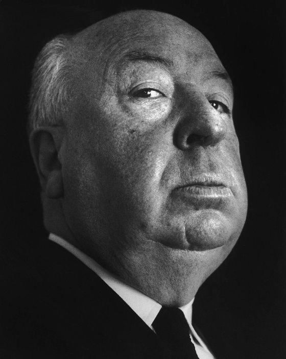
Photo Credit: Getty Images If someone is in a vegetative state is he or she aware of you? Even more interestingly, is he or she able to follow the plot of a suspense filled movie? Researchers at Western’s Brain and Mind Institute examined the brain activity of a man, who had been unresponsive for 16 years, while viewing a short Alfred Hitchcock film. Findings indicated that the man’s brain responded similarly to the brains of the participants in the control group. Medical Xpress discussed the study in a recent article:
Lorina Naci, a postdoctoral fellow from Western's Brain and Mind Institute, and her Western colleagues, Rhodri Cusack, Mimma Anello and Adrian Owen, reported their findings today in The Proceedings of the National Academy of Sciences (PNAS), in a study titled, A common neural code for similar conscious experiences in different individuals.
While inside the 3T Magnetic Resonance Imaging (MRI) Scanner at Western's Centre for Functional and Metabolic Mapping, participants watched a highly engaging short film by Alfred Hitchcock. Movie viewing elicited a common pattern of synchronized brainactivity. The long-time unresponsive participant's brain response during the same movie strongly resembled that of the healthy participants, suggesting not only that he was consciously aware, but also that he understood the movie.
"For the first time, we show that a patient with unknown levels of consciousness can monitor and analyze information from their environment, in the same way as healthy individuals," said Naci, lead researcher on the new study. "We already know that up to one in five of these patients are misdiagnosed as being unconscious and this new technique may reveal that that number is even higher."
Owen, the Canada Excellence Research Chair in Cognitive Neuroscience and Imaging, explained, "This approach can detect not only whether a patient is conscious, but also what that patient might be thinking. Thus, it has important practical and ethical implications for the patient's standard of care and quality of life."
The researchers hope that this novel method will enable better understanding of behaviorally unresponsive patients, who may be misdiagnosed as lacking consciousness.
Read the original article Here
- Comments (0)
- Single Dose of SSRIs Change Brain Connectivity After Only Three Hours
- By Jason von Stietz
- September 22, 2014
-

Photo Credit: Getty Images Could a single dose of antidepressants change the "architecture" of the brain? Researchers examined the effects of Selective Serotonin Reuptake Inhibitors (SSRI) on the brain using Magnetic Resonance Imaging (MRI). Although most patients do not see improvements in depression for weeks, the study found changes in brain connectivity only three hours after the dosage. These findings suggest that physicians might be able to use brain scans used to determine a patient's responsiveness to SSRIs. The study was recently discussed in the Los Angeles Times:
The findings could be a first step toward figuring out whether a relatively simple brain scan might one day help psychiatrists distinguish between those who respond to such drugs and those who don’t, an area of mystery and controversy in depression treatment.
Researchers at the Max Planck Institute in Leipzig, Germany, used a magnetic resonance imaging machine to compare connections in the gray matter of those who took SSRIs and those who did not. They were particularly interested in what goes on when the brain is doing nothing in particular.
They created 3-D maps of connections that “matter” to gray matter: interdependence, not just anatomical connection. They relied on a discovery in the late 1990s that low-frequency brain signaling during relative inactivity, such as daydreaming, is a good indicator of functional connectivity.
When more serotonin was available, this resting state functional connectivity decreased on a broad scale, the study found. This finding was not particularly surprising -- other studies have shown a similar effect in brain regions strongly associated with mood regulation.
But there was a two-fold shock: Some areas of the brain appeared to buck the trend and become more interdependent. And all the changes were evident only three hours after the single dosage.
“It was interesting to see two patterns that seemed to go in the opposite direction,” Sacher said. “What was really surprising was that the entire brain would light up after only three hours. We didn’t expect that.”
Most people who use antidepressants don't report any discernible change in mood for at least two weeks, said Sacher, who also is a psychiatrist.
The rapid connectivity shifts noted by the study might therefore be precursors to longer-term changes, perhaps starting with remodeling of synapses, the microscopic gaps where chemical neurotransmitters such as serotonin flood across to an adjacent brain cell, the study suggests. But this type of brain scanning can’t pick up changes at such a scale, so the hypothesis will have to be tested other ways, Sacher added.
More research also will be needed to explain why the functional connectivity of the cerebellum and thalamus apparently increased. The cerebellum, or “little brain” is a relatively primitive structure that processes signals from the spinal cord, relaying them to the thalamus and on up to the cortex. Many of those pathways are regulated by serotonin
Although the cerebellum is associated mostly with such basic functions as motor control and coordination, there are hints that it plays a role in higher cognitive tasks, perhaps by changing the way the thalamus relays signals to the cortex.
Although many open hypotheses remain to be explored, the brain maps produced by the study advance the effort to identify a brain “fingerprint” for those who might respond to SSRI therapy, researchers said.
Study subjects did not have diagnoses of depression, so researchers will need to generate similar maps among those diagnosed with depression, and re-map them during and after depressive episodes, as well as after treatment, Sacher said. Comparisons might then show whether a certain initial architecture predicts treatment success.
“In a perfect world, you would look not only at SSRIs, but all sorts of medications and non-pharmacological interventions,” Sacher said. “That would really help to tailor individual therapy for someone in the midst of a depressive episode.”
Read the original article Here
- Comments (0)
- The Adolescent Brain is Wired for Risk: Especially When Others are Looking
- By Jason von Stietz
- September 15, 2014
-

Photo Credit: NewsArt More than a billion dollars a year is spent in the United States each year educating teens on the dangers of drinking, smoking, drugs, reckless driver, and unsafe sex. However, risky behaviors among teens persist. In fact, illicit drug use among teens has increased over the past 20 years. Psychologists point to the still developing brain of adolescents for an explanation. Risky behavior among otherwise intelligent adolescents was discussed in a recent issue of The Atlanta Journal Constitution:
Adolescents are as smart as adults by their late teens; their memories are excellent, their reasoning skills sharp.
What they don’t have is the self-control and self-regulation of adults.
That is due, says Steinberg, to their developing brains. “Risk taking is a natural, hardwired and evolutionarily understandable feature of adolescence,” he writes. “Heightened risk-raking during adolescence is normal and, to some extent, inevitable.”
His book combines emerging brain science, parenting advice and political and education advocacy. Both his public policy and his parent recommendations are straightforward: Because it is hard to counter evolution and endocrinology, we ought to limit the opportunities for teens to exercise the immature judgment that comes with a developing brain.
In a telephone interview, Steinberg said, “I don’t want to send a message that we need to set our homes up as a kind of prison camp for kids. Kids need opportunities to be independent and have autonomy.”
“One reasonable example — given the fact we know a lot of experimenting with drugs or drinking or sex occurs in the afternoon hours when they are out of school and their parents are at work — is for schools to provide better after-school activities that have adult supervision,” he said. Funding for such programs could come from the budgets now underwriting ineffectual health education classes.
Steinberg encourages parents to require their teens participate in activities. “As a parent, you can say to your child, ‘You don’t have to do this specific activity, but you have to do something. You just can’t hang out with your friends. You can’t go over to Sally’s house because that’s the place where there are no parents.’ We know letting adolescents have a lot of unsupervised time in groups is a recipe for trouble. It’s not all kids. And it’s not all the time. But as a rule, it is true.”
There is no more vivid illustration of the impact of peers on teens than on the road. Letting teens drive with three friends in the car is akin to allowing them to drink and drive, said Steinberg. (Two recent accidents in which Georgia teens died, including a speed-related Murray County crash that killed four people, involved teens driving with friends. The Facebook page of the teen driver had a meme that reflects the role of peers. It read: "Good friends stop you from doing stupid stuff. Best friends do stupid stuff with you.")
Steinberg cites fascinating research on how teen behavior changes when kids have an audience of peers because the reward circuitry in their brains is activated. They are far more likely to drive fast, shoplift or drink with friends than when alone. In lab experiments, teens take more risks when told another teen is observing them on a computer screen.
While we often cast adolescence as something to be endured and survived, Steinberg says it also can be seen as an opportunity if we understand what teens need to thrive. And that is support, engagement, opportunities and limits. The best predictor of adolescent mental health, he says, is parent involvement.
Read the Full Article Here
- Comments (0)
- Recreational Marijuana Use Related to Brain Abnormalities
- By Jason von Stietz
- September 5, 2014
-

Photo Credit: Getty Images It is a commonly held belief that 'recreational" marijuana use is harmless. However, according to researchers at Harvard and Northwestern University, that may not be the case. A recent study found structural abnormalities in the nucleus accumbens and the amygdala of recreational marijuana users. News.Mic covered the story in a recent article:
According to a new study published in the Journal of Neuroscience, researchers from Harvard and Northwestern studied the brains of 18- to 25-year-olds, half of whom smoked pot recreationally and half of whom didn't. What they found was rather shocking: Even those who only smoked few times a week had significant brain abnormalities in the areas that control emotion and motivation.
"There is this general perspective out there that using marijuana recreationally is not a problem — that it is a safe drug," said Anne Blood, a co-author of the study. "We are seeing that this is not the case."
The science: Similar studies have found a correlation between heavy pot use and brain abnormalities, but this is the first study that has found the same link with recreational users. The 20 people in the "marijuana group" of the study smoked four times a week on average; seven only smoked once a week. Those in the control group did not smoke at all.
"We looked specifically at people who have no adverse impacts from marijuana — no problems with work, school, the law, relationships, no addiction issues," said Hans Breiter, another co-author of the study.
Using three different neuroimaging techniques, researchers then looked at the nucleus accumbens and the amygdala of the participants. These areas are responsible for gauging the benefit or loss of doing certain things, and providing feelings of reward for pleasurable activities such as food, sex and social interactions.
"This is a part of the brain that you absolutely never ever want to touch," said Breiter. "I don't want to say that these are magical parts of the brain — they are all important. But these are fundamental in terms of what people find pleasurable in the world and assessing that against the bad things."
Shockingly, every single person in the marijuana group, including those who only smoked once a week, had noticeable abnormalities, with the nucleus accumbens and the amygdala showing changes in density, volume and shape. Those who smoked more had more significant variations.
What will happen next? The study's co-authors admit that their sample size was small. Their plan now is to conduct a bigger study that not only looks at the brain abnormalities, but also relates them to functional outcomes. That would be a major and important step in this science because, as of now, the research indicates that marijuana use may cause alterations to the brain, but it's unclear what that might actually mean for users and their brains.
But for now, they are standing behind their findings.
"People think a little marijuana shouldn't cause a problem if someone is doing OK with work or school," said Breiter. "Our data directly says this is not so."
Read the Full Article Here
- Comments (0)


 Subscribe to our Feed via RSS