Blog
- fMRI Neurofeedback Used to Increase Confidence and Decrease Fear
- By Jason von Stietz, M.A.
- December 31, 2016
-

What does confidence look like in the brain? Is it possible to directly train confidence? Researchers at UCLA investigated the use of fMRI neurofeedback to increase confidence and decrease fear. Researchers decoded intricate patterns of brain activity associated with confidence and fear. Neurofeedback was then used to reward the brain activity patterns, which were unconscious to the participants. The study was discussed in a recent article in MedicalXpress:
The approach, developed by a UCLA-led team of neuroscientists, is described in two new papers, published in the journals Nature Communications and Nature Human Behaviour.
Their method could have implications for treating people with depression, dementia and anxiety disorders, including post-traumatic stress disorder, said Hakwan Lau, a UCLA associate professor of psychology and the senior author of both studies. It could also play a role in improving leadership training for executives and managers.
In the Nature Human Behaviour study, the researchers showed that they could reduce the brain's manifestation of fear using a procedure called decoded neurofeedback, which involves identifying complex patterns of brain activity linked to a specific memory, and then giving feedback to the subject—for example, in the form of a reward—based on their brain activity.
The researchers tested the technique on 17 undergraduate and graduate students in Japan. Participants were seated in a functional magnetic resonance imaging, or fMRI, scanner and shown patterns of vertical lines in four colors—red, green, blue and yellow. The blue and yellow images were always shown without shocks, but the red and green patterns were often accompanied by a small electrical shock administered to their feet.
As a result, the subjects' brain patterns began to register fear for the red and green images. But the scientists learned that they could use decoded neurofeedback to lessen the subjects' fear of the red pattern. They did this by giving the subjects a small cash reward—the equivalent of about 10 cents—each time they spontaneously thought about the red lines (but gave no rewards for thinking about the green lines), which the scientists could determine in real time based on their brain activity.
The following day, researchers tested whether the participants still had a fear response to the vertical lines. The red pattern, which had been frightening because it was paired with shocks, became less so because it now was paired with a positive outcome. With the reward as part of the equation, researchers found that participants perspired much less than when they had seen the red lines previously, and their brain's fear signal, centered in the amygdala, was significantly reduced.
"After just three days of training, we saw a significant reduction of fear," Lau said. "We changed the association of the 'fear object' from negative to positive."
Participants were not told what they had to do to earn the money—only that the reward was based on their brain activity and that they should try to earn as much money as possible. And each time participants were told they had won money, their brains demonstrated more of the same pattern that had just won them the cash reward.
Although participants tried to guess which of their thoughts were triggering the rewards—some guessed humming music or thinking about a girlfriend, for example—none actually figured out how they earned the money or recognized that the researchers had effectively reduced their fear of the red lines.
"Their brain activity was completely unconscious," Lau said. "That makes sense; a lot of our brain activity is unconscious."
Participants did still register fear on their fMRI scans when they saw the green pattern because, without the financial rewards, they still primarily associated the color with shocks.
The findings could help improve upon standard behavioral therapy, in which a person who is afraid of a certain object is exposed to photos of that object, or even the object itself—which can be frightening enough that many people cannot complete treatment. Lau said using "unconscious fear reduction," like in the experiment, could be more effective in many cases.
Instilling confidence
In the Nature Communications study, which was published today, Lau and his colleagues used decoded neurofeedback to increase people's confidence levels.
Ten participants were seated in an fMRI scanner and asked to watch a computer screen with hundreds of dots moving in different directions.
Participants were asked whether the majority of dots were moving to the left or the right, and how confident they were in their responses. That initial feedback gave the researchers a chance to see how high confidence and low confidence were represented in brain patterns.
Participants then were shown dots moving in random motion and told to think about anything—and that certain thoughts would earn them cash rewards. Every time the brain pattern looked like it was representing high confidence, the participant received a reward of up to the equivalent of 10 cents; subjects received smaller rewards if their brain activity indicated less confidence.
Next, the researchers showed the students the images from the first phase of the experiment—with numerous dots primarily moving in one direction or the other. The scientists found that although students weren't any better at guessing the primary direction of the dots' motion, they had become more confident in their guesses.
By studying brain patterns, Lau said, neuroscientists can decode people's thoughts about food, love, money and many other concepts, which eventually could help them design treatments for eating disorders, gambling addiction and more. The researchers next will determine whether the techniques described in the papers can be used to help patients with real phobias.
"We are cautiously optimistic," he said.
Read the original article Here
- Comments (0)
- Transcranial Magnetic Stimulation Reactivates Lost Memory
- By Jason von Stietz, M.A.
- December 19, 2016
-
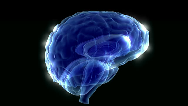
Photo Credit: Getty Images One’s ability to hold information in working memory is essential to daily functioning. Without working memory one would not be able to dial a phone number before forgetting it or remember the location of a chair in a room in order to avoid bumping into it. Previously, researchers believed that the brain needed to sustain elevated activity to maintain the memory, and once the activity ceased the memory was lost. However, researchers at University of Wisconsin Madison found that transcranial magnetic stimulation can retrieve memories once elevated brain activity has ceased. The study was discussed in a recent article in NeuroScientist News:
"A lot of mental illness is associated with the inability to choose what to think about," says Brad Postle, a psychology professor at the University of Wisconsin- (UW-) Madison. "What we're taking are first steps toward looking at the mechanisms that give us control over what we think about."
Postle's lab is challenging the idea that working memory remembers things through sustained brain activity. They caught brains tucking less-important information away somewhere beyond the reach of the tools that typically monitor brain activity—and then they snapped that information back into active attention with magnets.
Their latest study is published in the journal Science.
According to Postle, it's important to note that most people feel they are able to concentrate on a lot more than their working memory can actually hold. It's a bit like vision, in which it feels like we're seeing everything in our field of view, but details slip away unless you re-focus on them regularly.
"The notion that you're aware of everything all the time is a sort of illusion your consciousness creates," says Postle. "That is true for thinking, too. You have the impression that you're thinking of a lot of things at once, holding them all in your mind. But lots of research shows us you're probably only actually attending to—are conscious of in any given moment—just a very small number of things."
Postle's group conducted a series of experiments in which people were asked to remember two items representing different types of information (they used words, faces and directions of motion) because they'd be tested on their memories.
When the researchers gave their subjects a cue as to the type of question coming—a face, for example, instead of a word—the electrical activity and blood flow in the brain associated with the word memory disappeared. But if a second cue came letting the subject know they would now be asked about that word, the brain activity would jump back up to a level indicating it was the focus of attention.
"People have always thought neurons would have to keep firing to hold something in memory. Most models of the brain assume that," says Postle. "But we're watching people remember things almost perfectly without showing any of the activity that would come with a neuron firing. The fact that you're able to bring it back at all in this example proves it's not gone. It's just that we can't see evidence for its active retention in the brain."
The researchers were also able to bring the seemingly abandoned items back to mind without cueing their subjects. Using a technique called transcranial magnetic stimulation (TMS) to apply a focused electromagnetic field to a precise part of the brain involved in storing the word, they could trigger the sort of brain activity representative of focused attention.
Furthermore, if they cued their research subjects to focus on a face (causing brain activity associated with the word to drop off), a well-timed pulse of transcranial magnetic stimulation would snap the stowed memory back into attention, and prompt the subjects to incorrectly think that they had been cued to focus on the word.
"We think that memory is there, but not active," says Postle, whose work is supported by the National Institute of Mental Health. "More than just showing us it's there, the TMS can actually make that memory temporarily active again."
The study—conducted by Postle with Nathan Rose, a former UW-Madison postdoctoral researcher who is now a professor of psychology at the University of Notre Dame, and UW-Madison graduate students in psychology and neuroscience—suggests a state of memory apart from the spotlight attention of active working memory and the deep storage of more significant things in long-term memory.
"What's still unknown here is how the brain determines what falls away, and what enables you to retrieve things in the short-term if you need them," Postle says.
Studying how the brain apportions attention could eventually influence the way we understand and treat mental health disorders such as schizophrenia, in which patients focus on hallucinations instead of reality, and depression, which seems strongly related to spending an unhealthy amount of time dwelling on negative things.
"We are making some interesting progress with very basic research," says Postle. "But you can picture a point at which this work could help people control their attention, choose what they think about, and manage or overcome some very serious problems associated with a lack of control."
Read the original article here
- Comments (0)
- Spiritual Experience Studied Using fMRI
- By Jason von Stietz, M.A.
- December 9, 2016
-

Photo Credit: University of Utah Health Sciences Researchers from University of Utah as well as Harvard University investigated the brain’s response to a spiritual experience. The study utilized fMRI scans to measure the response of Mormon participants’ brains while viewing Mormon quotations and video of religious content. Participants reported experiencing feelings of peace and physical warmth. Findings indicated that areas of the brain related to focused attention, processing rewards, and moral reasoning. The study was discussed in a recent article in Medical Xpress:
Religious and spiritual experiences activate the brain reward circuits in much the same way as love, sex, gambling, drugs and music, report researchers at the University of Utah School of Medicine. The findings will be published Nov. 29 in the journal Social Neuroscience.
"We're just beginning to understand how the brain participates in experiences that believers interpret as spiritual, divine or transcendent," says senior author and neuroradiologist Jeff Anderson, M.D., Ph.D. "In the last few years, brain imaging technologies have matured in ways that are letting us approach questions that have been around for millennia."
Specifically, the investigators set out to determine which brain networks are involved in representing spiritual feelings in one group, devout Mormons, by creating an environment that triggered participants to "feel the Spirit." Identifying this feeling of peace and closeness with God in oneself and others is a critically important part of Mormons' lives—they make decisions based on these feelings; treat them as confirmation of doctrinal principles; and view them as a primary means of communication with the divine.
During fMRI scans, 19 young-adult church members—including seven females and 12 males—performed four tasks in response to content meant to evoke spiritual feelings. The hour-long exam included six minutes of rest; six minutes of audiovisual control (a video detailing their church's membership statistics); eight minutes of quotations by Mormon and world religious leaders; eight minutes of reading familiar passages from the Book of Mormon; 12 minutes of audiovisual stimuli (church-produced video of family and Biblical scenes, and other religiously evocative content); and another eight minutes of quotations.
During the initial quotations portion of the exam, participants—each a former full-time missionary—were shown a series of quotes, each followed by the question "Are you feeling the spirit?" Participants responded with answers ranging from "not feeling" to "very strongly feeling."
Researchers collected detailed assessments of the feelings of participants, who, almost universally, reported experiencing the kinds of feelings typical of an intense worship service. They described feelings of peace and physical sensations of warmth. Many were in tears by the end of the scan. In one experiment, participants pushed a button when they felt a peak spiritual feeling while watching church-produced stimuli.
"When our study participants were instructed to think about a savior, about being with their families for eternity, about their heavenly rewards, their brains and bodies physically responded," says lead author Michael Ferguson, Ph.D., who carried out the study as a bioengineering graduate student at the University of Utah.
Based on fMRI scans, the researchers found that powerful spiritual feelings were reproducibly associated with activation in the nucleus accumbens, a critical brain region for processing reward. Peak activity occurred about 1-3 seconds before participants pushed the button and was replicated in each of the four tasks. As participants were experiencing peak feelings, their hearts beat faster and their breathing deepened.
In addition to the brain's reward circuits, the researchers found that spiritual feelings were associated with the medial prefrontal cortex, which is a complex brain region that is activated by tasks involving valuation, judgment and moral reasoning. Spiritual feelings also activated brain regions associated with focused attention.
"Religious experience is perhaps the most influential part of how people make decisions that affect all of us, for good and for ill. Understanding what happens in the brain to contribute to those decisions is really important," says Anderson, noting that we don't yet know if believers of other religions would respond the same way. Work by others suggests that the brain responds quite differently to meditative and contemplative practices characteristic of some eastern religions, but so far little is known about the neuroscience of western spiritual practices.
The study is the first initiative of the Religious Brain Project, launched by a group of University of Utah researchers in 2014, which aims to understand how the brain operates in people with deep spiritual and religious beliefs.
Read the original article Here
- Comments (0)
 Subscribe to our Feed via RSS