Blog
- Poor Sleep Related to Biomarker for Alzheimer's
- By Jason von Stietz, PhD
- January 31, 2019
-
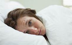
Getty Images According to the Alzheimer’s Association, 5.7 individuals in the United States are currently living with Alzheimer’s disease. Furthermore, it is the 6th leading cause of death in the United States. Researchers from the Washington University School of Medicine examined the relationship between sleep and biomarkers related to Alzheimer’s disease. The findings were discussed in a recent article in Neuroscience News and Research:
Poor sleep is a hallmark of Alzheimer’s disease. People with the disease tend to wake up tired, and their nights become even less refreshing as memory loss and other symptoms worsen. But how and why restless nights are linked to Alzheimer’s disease is not fully understood.
Now, researchers at Washington University School of Medicine in St. Louis may have uncovered part of the explanation. They found that older people who have less slow-wave sleep – the deep sleep you need to consolidate memories and wake up feeling refreshed – have higher levels of the brain protein tau. Elevated tau is a sign of Alzheimer’s disease and has been linked to brain damage and cognitive decline.
The findings, published Jan. 9 in Science Translational Medicine, suggest that poor-quality sleep in later life could be a red flag for deteriorating brain health.
“What’s interesting is that we saw this inverse relationship between decreased slow-wave sleep and more tau protein in people who were either cognitively normal or very mildly impaired, meaning that reduced slow-wave activity may be a marker for the transition between normal and impaired,” said first author Brendan Lucey, MD, an assistant professor of neurology and director of the Washington University Sleep Medicine Center. “Measuring how people sleep may be a noninvasive way to screen for Alzheimer’s disease before or just as people begin to develop problems with memory and thinking.”
The brain changes that lead to Alzheimer’s, a disease that affects an estimated 5.7 million Americans, start slowly and silently. Up to two decades before the characteristic symptoms of memory loss and confusion appear, amyloid beta protein begins to collect into plaques in the brain. Tangles of tau appear later, followed by atrophy of key brain areas. Only then do people start showing unmistakable signs of cognitive decline.
The challenge is finding people on track to develop Alzheimer’s before such brain changes undermine their ability to think clearly. For that, sleep may be a handy marker.
To better understand the link between sleep and Alzheimer’s disease, Lucey, along with David Holtzman, MD, the Andrew B. and Gretchen P. Jones Professor and head of the Department of Neurology, and colleagues studied 119 people 60 years of age or older who were recruited through the Charles F. and Joanne Knight Alzheimer’s Disease Research Center. Most – 80 percent – were cognitively normal, and the remainder were very mildly impaired.
The researchers monitored the participants’ sleep at home over the course of a normal week. Participants were given a portable EEG monitor that strapped to their foreheads to measure their brain waves as they slept, as well as a wristwatch-like sensor that tracks body movement. They also kept sleep logs, where they made note of both nighttime sleep sessions and daytime napping. Each participant produced at least two nights of data; some had as many as six.
The researchers also measured levels of amyloid beta and tau in the brain and in the cerebrospinal fluid that bathes the brain and spinal cord. Thirty-eight people underwent PET brain scans for the two proteins, and 104 people underwent spinal taps to provide cerebrospinal fluid for analysis. Twenty-seven did both.
After controlling for factors such as sex, age and movements while sleeping, the researchers found that decreased slow-wave sleep coincided with higher levels of tau in the brain and a higher tau-to-amyloid ratio in the cerebrospinal fluid.
“The key is that it wasn’t the total amount of sleep that was linked to tau, it was the slow-wave sleep, which reflects quality of sleep,” Lucey said. “The people with increased tau pathology were actually sleeping more at night and napping more in the day, but they weren’t getting as good quality sleep.”
If future research bears out their findings, sleep monitoring may be an easy and affordable way to screen earlier for Alzheimer’s disease, the researchers said. Daytime napping alone was significantly associated with high levels of tau, meaning that asking a simple question – How much do you nap during the day? – might help doctors identify people who could benefit from further testing.
“I don’t expect sleep monitoring to replace brain scans or cerebrospinal fluid analysis for identifying early signs of Alzheimer’s disease, but it could supplement them,” Lucey said. “It’s something that could be easily followed over time, and if someone’s sleep habits start changing, that could be a sign for doctors to take a closer look at what might be going on in their brains.”
View the original article here
- Comments (0)
- Smartphone App Reduces OCD Symptoms, Study Finds
- By Jason von Stietz, Ph.D.
- November 29, 2018
-
Researchers at the University of Cambridge developed a brain training app aimed at addressing obsessive compulsive disorder (OCD). The app, which can be used on a smart phone, involves people who suffer from OCD watching videos of themselves washing their hands, as excessive handwashing is a common symptom of OCD. The researchers tested the app in a study published in Scientific Reports. The study was discussed in a recent article in Medical Xpress:
In a study published in the journal Scientific Reports, Baland Jalal and Professor Barbara Sahakian from the Department of Psychiatry, show how just one week of training can lead to significant improvements.
One of the most common types of OCD, affecting up to 46% of OCD patients, is characterised by severe contamination fears and excessive washing behaviour. Excessive washing can be harmful as sometimes OCD patients use spirits, surface cleansers or even bleach to clean their hands. The behaviours can have a serious impact on people's lives, their mental health, their relationships and their ability to hold down jobs.
This repetitive and compulsive behaviour is also associated with 'cognitive rigidity' - in other words, an inability to adapt to new situations or new rules. Breaking out of compulsive habits, such as handwashing, requires cognitive flexibility so that the OCD patient can switch to new activities instead.
OCD is treated using a combination of medication such as Prozac and a form cognitive behavioural therapy ('talking therapy') termed 'exposure and response prevention'. This latter therapy often involves instructing OCD patients to touch contaminated surfaces, such as a toilet, but to refrain from then washing their hands.
These treatments are not particularly effective, however—as many as 40% of patients fail to show a good response to either treatment. This may be in part because often people with OCD have suffered for years prior to receiving a diagnosis and treatment. Another difficulty is that patients may fail to attend exposure and response prevention therapy as they find it too stressful to undertake.
For these reasons, Cambridge researchers developed a new treatment to help people with contamination fears and excessive washing. The intervention, which can be delivered through a smartphone app, involves patients watching videos of themselves washing their hands or touching fake contaminated surfaces.
Ninety-three healthy people who had indicated strong contamination fears as measured by high scores on the 'Padua Inventory Contamination Fear Subscale' participated in the study. The researchers used healthy volunteers rather than OCD patients in their study to ensure that the intervention did not potentially worsen symptoms.
The participants were divided into three groups: the first group watched videos on their smartphones of themselves washing their hands; the second group watched similar videos but of themselves touching fake contaminated surfaces; and the third, control group watched themselves making neutral hand movements on their smartphones.
After only one week of viewing their brief 30 second videos four times a day, participants from both of the first two groups—that is, those who had watched the hand washing video and those with the exposure and response prevention video—improved in terms of reductions in OCD symptoms and showed greater cognitive flexibilitycompared with the neutral control group. On average, participants in the first two groups saw their Yale-Brown Obsessive Compulsive Scale (YBOCS) scores improve by around 21%. YBOCS scores are the most widely used clinical assessments for assessing the severity of OCD.
Importantly, completion rates for the study were excellent—all participants completed the one week intervention, with participants viewing their video an average (mean) of 25 out of 28 times.
Mr Jalal said: "Participants told us that the smartphone washing app allowed them to easily engage in their daily activities. For example, one participant said 'if I am commuting on the bus and touch something contaminated and can't wash my hands for the next two hours, the app would be a sufficient substitute'."
Professor Sahakian said: "This technology will allow people to gain help at any time within the environment where they live or work, rather than having to wait for appointments. The use of smartphone videos allows the treatment to be personalised to the individual.
"These results while very exciting and encouraging, require further research, examining the use of these smartphone interventions in people with a diagnosis of OCD."
The smartphone app is not currently available for public use. Further research is required before the researchers can show conclusively that it is effective at helping patients with OCD.
Read the article Here
- Comments (0)
- MRI Shows Gray Matter Changes in those with Fibromyalgia Following CBT
- By Jason von Stietz, Ph.D.
- October 28, 2018
-

Getty Images People who suffer from fibromyalgia often suffer from both chronic pain and sleep disturbance, which can result in changes in atrophying of gray matter. Researchers recently conducted a piot study utilizing magnetic resonance imaging (MRI) to examine the relationship between cognitive behavior therapy and changes in gray matter in those with fibromyalgia. The findings were discussed in a recent article in Medical Xpress:
Results suggest that eight weeks of CBT-I can alter central nervous system structure in patients with fibromyalgia and insomnia. Individuals who received CBT-I demonstrated increases in cortical thickness following treatment, while individuals in a control groupshowed thinning of the cortex relative to baseline. Surprisingly, cognitive behavioral therapy for pain (CBT-P) failed to produce similar results as CBT-I.
"Our preliminary results suggest that while CBT-P seemed to merely reduce cortical atrophy, CBT-I produced increases in cortical thickness following treatment," said principal investigator Christina McCrae, Ph.D., professor in the department of psychiatry at the University of Missouri and director of the MizZzou Sleep Research Lab.
The study results are published in the Sept. 15 issue of the Journal of Clinical Sleep Medicine.
According to the authors, insomnia frequently occurs together with fibromyalgia. The Cognitive Activation Theory of Stress (CATS) posits that complaints such as pain and fatigue may result from a shared underlying psychobiological sensitization. Research using magnetic resonance imaging (MRI) also has demonstrated that fibromyalgia is associated with atrophy of cortical gray matter in certain brain regions.
This analysis used data from a larger clinical trial investigating the efficacy of CBT-I and CBT-P for fibromyalgia and chronic insomnia. From 2009 to 2012, participants were recruited from the community for the parent study, and a subset of participants also underwent MRI before and after eight weeks of treatment.
Thirty-seven patients were randomly assigned to CBT-I, CBT-P or a waiting list control group. Both interventions consisted of eight weekly, 50-minute individualized sessions with a trained therapist.
CBT-I consisted of sleep hygiene education, stimulus control, autogenic relaxation, sleep restriction, and cognitive therapy. CBT-P included pain education, progressive muscle relaxation, adaptive techniques for pacing activity, visual imagery relaxation, and cognitive restructuring.
The authors noted that these preliminary findings have clinical implications for people who have fibromyalgia.
"Demonstration that CBT-I, a relatively brief intervention, can reverse or resolve pain-related, maladaptive neural plasticity has important implications for chronic pain sufferers," McCrae said.
McCrae recently received a new research grant from the National Institute of Nursing Research (NINR) to study the treatment of chronic pain using CBT-I. The trial will explore McCrae's idea that improving sleep in women with fibromyalgia will also improve their pain by promoting positive changes in how the brain processes and responds to pain.
View the original article Here
- Comments (0)
- Vagus Nerve Essential to Brain's Reward and Motivation System
- By Jason von Stietz, PhD
- September 30, 2018
-
Caption Researchers at the Icahn School of Medicine at Mount Sinai examined the relationship between the vagus nerve and the brain’s reward and motivation system. Findings of the study could assist in the future efforts for vagal stimulation therapy. The study was discussed in a recent article in Medical Xpress:
Previous research established the gut as a major regulator of motivational and emotional states but until now, the relevant gut-brain neuronal circuitry remained elusive. The vagus nerve, the longest of the cranial nerves, contains motor and sensory fibers and passes through the neck and thorax to the abdomen. Traditionally, scientists believed that the nerve exclusively mediated suppressive functions such as fullness and nausea; in contrast, circulating hormones, rather than vagal transmission, were thought to convey reward signals from the gut to the brain.
"Our study reveals, for the first time, the existence of a neuronal population of 'reward neurons' amid the sensory cells of the right branch of the vagus nerve," says Ivan de Araujo, DPhil, Senior Faculty in the Department of Neuroscience at the Icahn School of Medicine at Mount Sinai and senior author of the paper. "We focused on challenging the traditional view that the vagus nerve is unrelated to motivation and pleasure and we found that stimulation of the nerve, specifically its upper gut branch, is sufficient to strongly excite reward neurons lying deep inside the brain."
The branches of the vagus nerve are intricately intermingled, making it extremely difficult to manipulate each organ separately. To address this challenge, the research team employed a combination of virally delivered molecular tools that allowed them to exclusively target the vagal sensory neurons connected to the stomach and upper intestine.
Specifically, researchers combined different viruses carrying molecular tools in a way that allowed them to optically activate vagal neurons connected to the gut while vagal neurons leading to other organs remained mute. The approach, a state-of-the-art technique known as "optogenetics," allows investigators to use light to manipulate the activity of a prespecified set of neurons.
The study revealed that the newly identified reward neurons of the right vagus nerve operate under the same constraints attributed to reward neurons of the central nervous system, meaning they link peripheral sensory cells to the previously mapped populations of reward neurons in the brain. Strikingly, neurons of the left vagus were associated with satiety, but not with reward. The research team's anatomical studies also revealed, for the first time, that the right and left vagal branches ascend asymmetrically into the central nervous system.
"We were surprised to learn that only the right vagal branch eventually contacts the dopamine-containing reward neurons in the brainstem," explained Wenfei Han, MD, Ph.D., Assistant Professor of Neuroscience at the Icahn School of Medicine at Mount Sinai and lead author of the study. Dopamine is a neural transmitter known to be essential for reward and motivation.
The uncovering of right gastrointestinal vagal neurons as conveyors of reward signals to the brain opens opportunities for novel, more specific stimulation targets that may increase the efficacy of vagal nerve stimulation therapy, a treatment that involves delivering electrical impulses to the vagus nerve, for patients suffering from emotional and eating disorders.
Read the original article Here
- Comments (0)
- Transcranial Magnetic Stimulation Device FDA Approved for OCD Treatment
- By Jason von Stietz, PhD
- August 31, 2018
-

According to the DSM 5, the international prevalence rate of obsessive compulsive disorder (OCD) is about 1% to 2%. OCD involves recurrent and persistent unwanted thoughts, which individuals attempts to suppress through repetitive compulsive behaviors. Depending on the severity, OCD can drastically impede an individual’s daily functioning. Fortunately, patients who have not responded to traditional treatment now have greater access to a cutting-edge treatment called transcranial magnetic stimulation (TMS), as a TMS device was recently approved by the FDA. This news was recently discussed in an article in Medical Xpress:
Transcranial magnetic stimulation uses magnetic fields to stimulate nerve cells in the brain. The FDA approved it as a treatment for major depression in 2008 and for treating pain associated with certain migraines in 2013.
"Transcranial magnetic stimulation has shown its potential to help patients suffering from depression and headaches," said Carlos Pena, director of the division of neurological and physical medicine devices at the FDA's Center for Devices and Radiological Health.
"With today's marketing authorization, patients with OCD who have not responded to traditional treatments now have another option," Pena added in an agency news release.
The approval of the Brainsway Deep Transcranial Magnetic Stimulation System was based on a study of 100 OCD patients. Thirty-eight percent of those treated with the device had a more than 30 percent reduction in the severity of their symptoms, compared with 11 percent of those treated with a non-working ("sham") device.
No serious reactions to treatment with the device were reported, according to the FDA.
People with OCD have uncontrollable, reoccurring thoughts and behaviors. About 1 percent of U.S. adults had OCD in the past year, according to the U.S. National Institute of Mental Health. The disorder is typically treated with medication, psychotherapy or both. Most patients respond to treatment, but some continue to have symptoms.
View the original article Here
- Comments (0)
- Heatwave Impacts Cognitive Ability in College Students
- By Jason von Stietz, Ph.D.
- July 28, 2018
-

People often assume that young and healthy individuals are largely unaffected by a heatwave. People also tend to focus on the dangers of exposure to the heat outside and neglect the possible dangers of high temperatures in buildings without air conditioning. Researchers from Harvard T.H. Chan School of Public Health studied the effects of college students in un-air-conditioned dormitories versus their counterparts living in air-conditioned dormitories. Those living without air-conditioning during a heatwave had slower reaction times and scored less accurately on cognitive tests. The study was discussed in a recent article in Medical Xpress:
"Most of the research on the health effects of heat has been done in vulnerable populations, such as the elderly, creating the perception that the general population is not at risk from heat waves," said Jose Guillermo Cedeño-Laurent, research fellow at Harvard Chan School and lead author of the study. "To address this blind spot, we studied healthy students living in dorms as a natural intervention during a heat wave in Boston. Knowing what the risks are across different populations is critical considering that in many cities, such as Boston, the number of heat waves is projected to increase due to climate change."
The study will be published online July 10, 2018 in PLOS Medicine as part of a special issue dedicated to climate change and health.
Extreme heat can have severe consequences for public health and is the leading cause of death of all meteorological phenomena in the U.S. Temperatures around the world are rising, with 2016 marking the warmest year on record for the past two centuries. While the health impacts of extreme heat are well documented, most studies to date have focused on vulnerable populations, including the very young or the elderly, and tend to be epidemiologic studies that use outdoor temperature records. Understanding the effects of indoor temperatures is important given that adults in the U.S. spend 90% of their time indoors.
For this new study, researchers tracked 44 students in their late teens and early 20s living in dorm rooms. Twenty-four of the students lived in adjacent six-story buildings that were built in the early 1990s and had central AC. The remaining 20 students lived in low-rise buildings constructed between 1930 and 1950 that did not have AC. Researchers outfitted each student's room with a device that measured temperature, carbon dioxide levels, humidity, and noise levels, and tracked their physical activity and sleep patterns with wearable devices.
The study was conducted over 12 consecutive days in the summer of 2016. The first five days consisted of seasonable temperatures, followed by a five-day-long heat wave, and then a two-day cooldown. Each day the students took two cognition tests on their smartphones right after waking up. The first test required students to correctly identify the color of displayed words and was used to evaluate cognitive speed and inhibitory control—or the ability to focus on relevant stimuli when irrelevant stimuli are also present. The second test consisted of basic arithmetic questions and was used to assess cognitive speed and working memory.
The findings showed that during the heat wave, students in the buildings without AC performed worse on the tests than students in the air-conditioned dormitories and experienced decreases across five measures of cognitive function, including reaction times and working memory. During the heat wave, students in buildings without AC experienced 13.4% longer reaction times on color-word tests, and 13.3% lower addition/subtraction test scores compared with students with air-conditioned rooms. Combined, these data show that students in rooms with AC were not just faster in their responses, but also more accurate.
Interestingly, the most significant difference in cognitive function between the two groups was seen during the cooldown period, when outdoor temperatures began to subside but indoor temperatures remained elevated in the dormitories without air conditioning.
"Indoor temperatures often continue to rise even after outdoor temperatures subside, giving the false impression that the hazard has passed, when in fact the 'indoor heat wave' continues," said Joseph Allen, assistant professor of exposure assessment science and co-director of the Center for Climate, Health, and the Global Environment (C-CHANGE) at Harvard Chan School and one of the study's senior authors. "In regions of the world with predominantly cold climates, buildings were designed to retain heat. These buildings have a hard time shedding heat during hotter summer days created by the changing climate, giving rise to indoor heat waves."
View the original article Here
- Comments (0)
- Cognitive Training Reduces Depression
- By Jason von Stietz, M.A.
- June 30, 2018
-
.jpg)
iStock About half of individuals who suffer a traumatic brain injury (TBI) develop depression within a year. However, would a treatment designed to address cognitive symptoms also reduce depression? Researchers from the Center for Brain Health at the University of Texas at Dallas examined the use of cognitive training programs to address depression in those who suffered a TBI. Findings indicated that cognitive training, even when not directed toward psychiatric symptoms, related to not only cognitive improvements but also reductions in depression. The study was discussed in a recent article in Medical Xpress:
The recent study, published in Human Brain Mapping, revealed significant reductions in the severity of depressive symptoms, increased ability to regulate emotions, increases in cortical thickness and recovery from abnormal neural network connectivity after cognitive training.
"To our knowledge, this is the first study to report brain change associated with reduced depression symptoms after cognitive training," said Dr. Kihwan Han, a research scientist at the Center for BrainHealth who works in the lab of Dr. Daniel Krawczyk. Han is the lead author of the study.
"Overall, these findings suggest that cognitive training can reduce depressive symptoms in patients with traumatic brain injury even when the training does not directly target psychiatric symptoms," he said.
A past study involving the same protocol showed cognitive gains as well as similar changes in cortical thickness and neural network connectivity.
This study included 79 individuals with chronic TBI who all were at least six months post-injury. These individuals were randomly assigned into one of two groups: strategy-based training, which used the Strategic Memory Advanced Reasoning Training (SMART) program developed at the center; and information-based training, which used the Brain Health Workshop program. Researchers used the Beck Depressive Inventory to classify 53 of the participants as depressed.
The participants' depressive-symptom severity, psychological functioning scores and data from magnetic resonance imaging brain scans were collected before training, after training and three months post-training. Scans were used to study changes in brain structure and neural network connectivity.
Both training programs consisted of 12 one-and-a half-hour sessions over eight weeks that included quizzes, homework assignments, and projects conducted in small group settings that involved social interactions.
All participants in the depressed group showed significantly reduced depressive symptoms that were associated with improvements in cognitive and daily life functioning. According to Han, the social engagements, cognitive stimulation from new learning opportunities and hope of improvement afforded by both programs may help explain the reductions in depressive symptoms.
Based on the observed brain change patterns, Han suggested that improved emotion regulation also may be related to the reduced depressive symptoms. Over time, the reductions in depression symptom severity correlated with increased cortical thickness within the prefrontal cortex—an area of the brain responsible for executive functions needed for emotional control—and reductions in abnormally elevated neural connectivity within this region.
"Identifying what changes are happening in the brain when interventions successfully reduce depressive symptoms could allow us to create more effective, pharmaceutical-free approaches to help alleviate depression in people who experience chronic traumatic brain injury symptoms," said study author Dr. Sandra Bond Chapman, founder and chief director of the Center for BrainHealth, and Dee Wyly Distinguished University Professor.
Read the original article Here
- Comments (0)
- Is Mindfulness Different Than Relaxation?
- By Jason von Stietz, M.A.
- June 21, 2018
-

Are the effects of mindfulness meditation the same as relaxation training? Mindfulness meditation often leads to feelings of calmness. However, the goal of mindfulness is to practice non-judgmental present moment awareness and not necessarily. Does this difference in goals lead to changes in the brain’s response? Researchers at Massachusetts General Hospital led a study utilizing functional magnetic resonance imaging to examine the difference in neural activity of participants engaging in mindfulness based stress reduction versus relaxation response training. The study was discussed in a recent article in MedicalXpress:
There are two widely used meditation-based stress reduction courses. One is based on the relaxation response—first described by Herb Benson, MD, director emeritus of the MGH-based Benson-Henry Institute for Mind Body Medicine - which focuses on eliciting a physiologic state of deep rest, the opposite of the "fight or flight" stress response. The other is Mindfulness-Based Stress Reduction, developed by Jon Kabat-Zinn, Ph.D., of the University of Massachusetts Medical School, which emphasizes a particular, non-judgmental attitude termed "mindfulness" as key to stress reduction. Although both interventions are based on meditation, the scientific philosophies and meditative traditions upon which each is founded are different, and these differences are reflected in the instructions and exercises taught to patients.
"If the hypotheses proposed by the programs' creators are in fact correct, they imply that these programs promote wellness through different mechanisms of action," says Sara Lazar, Ph.D., of the MGH Psychiatric Neuroscience Research Program, senior author of the current report and assistant professor of Psychology at Harvard Medical School. "Such a finding would suggest that these programs could potentially have different effects on disease."
To investigate that possibility, healthy adults with high levels of stress were randomized to two 8-week programs—18 completed the relaxation response program, and 16 completed the mindfulness program. Both programs successfully decreased stress and increased mindfulness in participants. However, the mindfulness program resulted in further improvements in measures such as self-compassion and rumination, clearly indicating that the programs are not the same, Lazar says.
To further understand the similarities and differences between the programs, the team measured brain activity during a meditation technique common to both programs—a body scan, in which attention is moved sequentially throughout the body to develop bodily awareness. While the relaxation response program instructs participants to deliberately relax each body area as they become aware of it, the mindfulness program just emphasizes mindful awareness and acceptance "without any attempt to change anything."
Lead author Gunes Sevinc, Ph.D., a research fellow in Lazar's laboratory says, "By directly comparing the body-scan meditations, which differed only in cognitive strategy, we were able to identify the brain regions that are involved in mediating the common and differential strategies employed by each intervention."
The results showed that the strength of neural interaction between brain regions associated with present-moment awareness and bodily attention increased during both types of body-scan meditation. But each program also showed unique patterns of brain activity in line with the different theoretical orientation of each program. The relaxation response body scan strengthened coupling between neural regions commonly associated with deliberate control, including inferior frontal gyrus and supplementary motor areas. Conversely, the mindfulness body scan strengthened coupling between neural regions associated with sensory awareness and perception, including the insula and the pregenual anterior cingulate.
"These findings indicate that the programs are working through different neural mechanisms," says Sevinc, "The relaxation response program is working more through deliberate control mechanisms, while the mindfulness program is working more through sensory awareness mechanisms. It is somewhat analogous to weight training vs. aerobic exercise—both are beneficial, but each has its unique mechanism and contribution."
Norman Farb, Ph.D., of the University of Toronto Department of Psychology, who was not part of the study, says, "Professor Lazar's neuroimaging study helps us to better appreciate how these seemingly similar practices differ in important ways. Both practices seem to promote access to neural representations of the body, but they differ in how such representations are structured. This study is important for beginning to inform the public about key differences between conceptually similar therapeutic approaches, which may in turn allow people to make more skillful decisions about which practice might be right for their personal improvement."
Lazar notes that future studies will be needed to determine whether these neural and psychological differences impact specific diseases in unique ways.
Read the original article Here
- Comments (0)
- Does Music Reduce Anxiety in Individuals with Alzheimer's?
- By Jason von Stietz, M.A.
- May 31, 2018
-
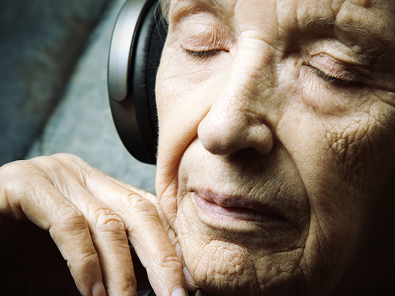
Why is it that we get chills when we listen to music? Why do our favorite old songs bring us right back to certain times in our life? In fact, this effect is seen even in those with Alzheimer’s disease. Researchers at the University of Utah Health examined the use of music in managing anxiety in individuals suffering from Anxiety. The study was discussed in a recent article in MedicalXpress:
"People with dementia are confronted by a world that is unfamiliar to them, which causes disorientation and anxiety" said Jeff Anderson, M.D., Ph.D., associate professor in Radiology at U of U Health and contributing author on the study. "We believe music will tap into the salience network of the brain that is still relatively functioning."
Previous work demonstrated the effect of a personalized music program on mood for dementia patients. This study set out to examine a mechanism that activates the attentional network in the salience region of the brain. The results offer a new way to approach anxiety, depression and agitation in patients with dementia. Activation of neighboring regions of the brain may also offer opportunities to delay the continued decline caused by the disease.
For three weeks, the researchers helped participants select meaningful songs and trained the patient and caregiver on how to use a portable media player loaded with the self-selected collection of music.
"When you put headphones on dementia patients and play familiar music, they come alive," said Jace King, a graduate student in the Brain Network Lab and first author on the paper. "Music is like an anchor, grounding the patient back in reality."
Using a functional MRI, the researchers scanned the patients to image the regions of the brain that lit up when they listened to 20-second clips of music versus silence. The researchers played eight clips of music from the patient's music collection, eight clips of the same music played in reverse and eight blocks of silence. The researchers compared the images from each scan.
The researchers found that music activates the brain, causing whole regions to communicate. By listening to the personal soundtrack, the visual network, the salience network, the executive network and the cerebellar and corticocerebellar network pairs all showed significantly higher functional connectivity.
"This is objective evidence from brain imaging that shows personally meaningful music is an alternative route for communicating with patients who have Alzheimer's disease," said Norman Foster, M.D., Director of the Center for Alzheimer's Care at U of U Health and senior author on the paper. "Language and visual memory pathways are damaged early as the disease progresses, but personalized music programs can activate the brain, especially for patients who are losing contact with their environment."
However, these results are by no means conclusive. The researchers note the small sample size (17 participants) for this study. In addition, the study only included a single imaging session for each patient. It is remains unclear whether the effects identified in this study persist beyond a brief period of stimulation or whether other areas of memory or mood are enhanced by changes in neural activation and connectivity for the long term.
"In our society, the diagnoses of dementia are snowballing and are taxing resources to the max," Anderson said. "No one says playing music will be a cure for Alzheimer's disease, but it might make the symptoms more manageable, decrease the cost of care and improve a patient's quality of life."
Read the original article Here
- Comments (0)
- Brain Science International is pleased to introduce the addition of Dr. J. Lucas Koberda, M.D., Ph.D. to our panel of experts
- By Jason von Stietz, Ph.D.
- April 3, 2018
-
Brain Science International is pleased to introduce the addition of Dr. J. Lucas Koberda, M.D., Ph.D. to our panel of experts. Dr. Koberda will be reading EEG’s and producing qEEG reports as well as neurofeedback recommendations.
Dr. J. Lucas Koberda, M.D.,Ph.D.

Dr. J. Lucas Koberda is a board certified neurologist and an internationally trained physician who completed his residency in Neurology at the Oregon Health Sciences University in Portland, Oregon. Prior to his neurological training, he received his Ph.D. based on his research in the area of tumor immunology. His research skills were also enhanced by post-doctoral training completed at MD. Anderson Cancer Center in Houston, Tx. Dr. Koberda is currently affiliated with The Florida State University College of Medicine. Dr. Koberda also has founded and became CEO of "Brain Enhancement, Inc." non-for profit organization. This organization focuses on neuromodulation and modern therapy of many neuropsychiatric disorders including Alzheimer's dementia, TBI, depression/anxiety, autistic spectrum/ADHD, epilepsy and chronic pain.
His main interest is in neuro-psychiatry and cognitive enhancement. He uses the newest technology of QEEG and Neurofeedback to successfully diagnose and treat many medical conditions including seizures,
headaches, fibromyalgia chronic pain, anxiety, depression, and prior stroke. Dr. Koberda has also effectively introduced neurofeedback protocols for a cognitive enhancement which may help students and professionals to improve their memory, concentration, verbal function or information processing speed.
Dr. Koberda also has been appointed in August, 2010 by Governor Charlie Christ as a member of the Alzheimer’s Disease Advisory Committee for a 4 years term (The Committee serves as an advising body to the Florida State Government). Dr. Koberda has published multiple publications in different scientific journals and serves as international neuroscience speaker and consultant.
Dr. Koberda has been appointed to the Editorial Board of the "Journal of Neurology and Stroke", "International Journal of Emergency Mental Health and Human Resilience", "Journal of Neurology and Neurobiology" and "Journal of Psychology and Clinical Psychiatry". He also serves as a reviewer-"Clinical EEG and Neuroscience" and the above journals.
Clinical skills and interests: EEG/QEEG/LORETA/Electrical Imaging (Brain Mapping), Neurofeedback, Neurotherapy, Magnetic and Electrical Stimulation, Neuromodulation, Neuropsychiatry, Forensic- neurology, EMG-NCV, Neuro-rehabilitation, IME, Teaching, Consultations, Academic and Research Interest, Clinical Trials, Public Health, Tele-neurology.
Director and Owner- J. Lucas Koberda, MD, PhD, Neurology, PL, Tallahassee Neurobalance Center.
www.TallahasseeNeuroBalanceCenter.com
Founder and CEO-Brain Enhancement, Inc.- non-for profit organization-since December, 2014.
Florida State University-Neurologist at FSU Health and Wellness Center-960 Learning Way, Tallahassee,FL 32306-since November, 2016.
Active and Recent Hospital Privileges:
Neurology Consultant-Florida State Psychiatric Hospital, Chattahoochee, FL-2007-December, 2014. Consulting Neurologist- HealthSouth-Rehabilitation Hospital, Tallahassee, FL.-2007- February, 2015. CRMC Hospital-Tallahassee, FL-community privileges.
Government Appointments:
Alzheimer Disease Advisory Committee (Florida)-appointed by the Florida Governor- Charlie Christ- August 2010-October 2012.
Licensure - Active: Florida, New York State, Utah, Georgia
Academic Association:
2012-Professor of Neurology-Carrick Institute for Graduate Studies, Cape Canaveral, FL.
2007-Clinical Assistant Professor of Neurology-Florida State University-College of Medicine, Tallahassee, FL
2001-Clinical Assistant Professor of Neurology-Albany Medical College, Albany, New York.Neurology and Geriatrics Elective Teaching Faculty 2010-2011.
Neurology Elective Director-Florida State University-College of Medicine-CRMC hospital-2007-2010.International Boards:
2013- 2014-Member of the International Board of Quantitative Electrophysiology (IBQE)
Editorial Board:
International Journal of Emergency Mental Health and Human Resilience-since 2014 Journal of Psychology and Clinical Psychiatry-since 2014,
Journal of Neurology and Stroke-since 2014,
Journal of Neurology and Neurobiology-since 2014,Valetudinaria-Progress in Clinical and Military Medicine (European medical journal)-2005-2013.
Journal Reviewer:
Clinical EEG and Neuroscience-since 2013
Journal of Psychology and Clinical Psychiatry-since 2014.
Journal of Neurology and Stroke-since 2014
International Journal of Emergency Mental health and Human Resilience-since 2014Foreign languages: English and Polish-Fluently, Russian and German-basic.
Education and Training:
Undergraduate: 1976 - 1980 M. Kopernik School Gdansk, Poland
Graduate: 1980 - 1986 Medical School in Gdansk-graduation-Oct 24, 1986 M. D.
Postgraduate Training and Employment:
Nov 1, 1986- Sep 30, 1989
Summer 1988-
Internal Medicine Residency A. Hellmann, M.D.-
Director Training, Medical School in Gdansk, Gdansk, PolandVisiting Scientist Prof. Castoldi, M.D.-Director Department of Hematology- University of Ferrara, Italy
Oct 1, 1989-
Sep 30, 1990
Hematology, J.W. Goethe University, Frankfurt/M., GermanyTX
Postdoctoral Training L. Bergmann, M.D.-Director Department of
Oct 25, 1990- Postdoctoral Training R. Moser, M. D. -Director
Sept 30, 1991 Department of Neurosurgry E.A. Grimm, Ph.D.-DirectorAnd Department of Tumor Biology UT M. D. Anderson Cancer Center, Houston,
Oct 1, 1991- Postdoctoral Training R.E. Champlin, M.D.-Director
April 24, 1994 Dept. of Hematology-BMT Section, UT M. D. Anderson Cancer Center, Houston, TXRelocation to Pennsylvania
July 1, 1994- June 30, 1995
July 1, 1995- June 30, 1998
Oct. 26, 1995 entitled
Transitional Resident-PGY-1, Easton Hospital, Easton, PA 18042 K.H Wildrick, M.D.-Director
Neurology Resident-Oregon Health Sciences University, Portland R.H. Whitham, M.D.-Director
Ph.D., degree in Immunology and Experimental Tumor Immunotherapy-paper
“Evaluation of oncolytic potential of human lymphocytes activated with low dose of IL-2 and anti-CD3 and
Director-Prof. Jedrzejczak.
July 1, 1998- July 2006 March, 2004- March 2005
August 1, 2006-
Solo Neurology Practice in Potsdam, NY
Medical Director of Acute Rehabilitation Unit – CHMC-Ogdensburg, NYpresent- Neurology practice in Tallahassee, FL
anti-CD28 antibodies.”-Graduated from Medical School in Warsaw-
2007-2010-Capital Regional Medical Center-Co-director of Stroke program and Neurology Elective Director-Florida State University-College of Medicine-CRMC hospital
May, 2000-2020 Board certification-Neurology- by the American Board of Psychiatry and Neurology Society-Organization Memberships:
1996 - American Academy of Neurology, Active Member-Epilepsy and Stroke section
1999-2004- Rotary International2003-Vice President-St. Lawrence County Medical Society, NY
2004-2006 President - St. Lawrence County Medical Society, NY
2006- Member of Rural and Preventive Medicine Committee –MSSNY
2006-Delegate from St. Lawrence County to House of Delegate meeting in Buffalo, NY2006-Member of Committee for Governmental Affairs B
2007-2012-Board of Directors-Epilepsy Association of the Big Bend, Tallahassee, FL. 2011-The Society of Applied Neurosciences.
2012-International Society for Neurofeedback and Research
2012-The Biofeedback Society of Florida.
2013-EEG and Clinical Neuroscience Society (ECNS).Medical Advocacy and Advising:
1. May-2004- Neurology on the Hill (Washington DC) - advocacy for development of stroke centers, malpractice reforms, Medicare – organized by American Academy of Neurology (AAN).
2. June-2005- Neurology on the Hill- advocacy for malpractice reforms, Medicare and Medicaid reforms- organized by AAN.
3. Spring 2005 - Albany NY-Advocacy on the State level- Malpractice reforms, access of Medicaid patient to care and services.
4. 2003-2004- Advocacy against obesity in children- resulted in elimination of sodas and “junk” food from local schools
5. 2004-2005-Advocacy in support of “self-pay” patients and insurance coverage.
6. May-2006- Neurology on the Hill- advocacy for fixing Medicare payment formula-organized by AAN.7. 2006- Health advisory meeting-to Congressman John M. McHugh (23rd. Congressional District of State of New York)-currently 21st United States Secretary of the Army (in the President Obama’s Cabinet).
7. March 2008-Advocacy to restrict trade medication substitution by generic anti-seizure medications without physician’s and patient’s consent-Tallahassee, FL
8. December 22nd, 2008-Host and moderator-Tallahassee Health Care Forum-the community discussion regarding health care reforms.
9. April 16th, 2009-organizer and moderator of the fundraiser to fight high infant mortality rate in Leon County, Tallahassee FL
10. October 8th, 2009-Organizer of fundraising and silent auction to support under-funded Tallahassee institutions including Epilepsy Association of the Big Bend and Neighborhood Health Services, Inc - serving underprivileged population with no health insurance.
Selected and Invited Presentations:
1999-Application of neurophysiologic testing for neurological diagnosis- Clarkson University Health Sciences- Potsdam, NY.
1999- Traumatic Brain Injury- Clarkson University Health Sciences- Potsdam, NY.
2004- New options in the treatment of epilepsy- Claxton-Hepburn Medical Center, Ogdensburg, NY.
2005- Introduction to Neurology- St. Lawrence University, Canton, NY.
2006- Introduction to Neurology- Florida State University, Tallahassee FL
2008-Introduction to diagnosis and treatment of epilepsy, Florida State Psychiatric Hospital, Tallahassee FL
2/2011-Current options in the treatment of epilepsy-hosted by the Epilepsy Association of The Big Bend.
10/2011-Quantitative EEG and Brain Disorders-invited speaker during the Brain Symposium in Tallahassee, FL.
5/25/2012-QEEG and Neurofeedback with Z-score LORETA in neurological practice-multiple-cases presentation-Cancun, Mexico-Z-score Neurofeedback workshop.
6/4/2012-Application of quantitative EEG in epilepsy and other neurological disorders-invited for the Grand Rounds presentation by the Neurology Department (Epilepsy Section) Medical University of Southern Carolina-Charleston, SC.
7/11/2012-QEEG and Neurofeedback in clinical practice-Grand Rounds presentation-Invited by Tallahassee Memorial Hospital in Tallahassee- invited by the residency program director.
7/21/2012—QEEG assessment with Z-score & LORETA neurofeedback in neuro-psychiatric disorders- invited speaker for a workshop presentation during The Biofeedback Society of Florida Meeting in Orlando, FL.
3/26/2013-Electrical Neuroimaging and Neurofeedback-Rotary Club, Tallahassee, FL.
8/8/2013-Electrical Imaging in Neuro-psychiarty-invited by the Tallahassee Memorial Hospital program director of Internal Medicine Residency Training-combined FSU/TMH hospital, Tallahassee, FL.
8/30/2013-The role of LORETA Z-score Neurofeedback in the Treatment of Neuropsychiatric Disorders- invited by Dr. Georges Otte-Medical Director of Neuropsychiatric Hospital-Ghent, Belgium.
9/21/2013-LORETA Z-score Neurofeedback in Neuropsychiatry-ISNR meeting in Dallas, TX-presented during the small group discussion section.
11/2/2013-“Z-score LORETA Neurofeedback in Clinical Practice”-invited speaker lecture during the Southeastern Biofeedback & Clinical Neuroscience Association conference in Lake Junalusca, NC.
1/7/2014-“Application of QEEG (Brain Mapping) in psychiatry-clinical implication for diagnosis and treatment”-TMH-psychiatry department, Tallahassee, FL.
3/20/2014-“Electrical Brain Imaging Cognitive Enhancement with LORETA Z-score Neurofeedback”- Advanced Psychology lecture-L. Chiles High School-Tallahassee, FL.
7/29/2014-“Electrical Brain Imaging LORETA Z-score Neurofeedback”-invited presentation-medical students-FSU.
8/15/2014-“Enhance Your Brain”-invited presentation at Maguire Center-Westminster Oaks, Tallahassee, FL.
2/12/2015-“QEEG/LORETA/Electrical imaging and Z-score LORETA Neurofeedback in neuropsychiatric diagnosis and therapy-presented during “Real-time Functional Imaging and Neurofeedback” international conference organized by University of Florida in Gainesville, FL.
2/21/2015-“Cognitive enhancement using LORETA Z-score Neurofeedback-presented during 30th Annual Alzheimer’s Disease Education and Training Conference-FSU College of Medicine-Tallahassee, FL.
2/2015-“QEEG and neurofeedback in Autistic Spectrum disorders”-invited presentation-by CARDS-FSU Autism Institute-invited by Dr. Amy Whetherby.
12-11-2015-“Neuromodulation Therapy of TBI, Cognitive Problems and Dementia”-Keynote invited speaker during the international conference-International Symposium on Clinical Neuroscience-Orlando, FL.
8/26/2016-Invited keynote speaker-QEEG and LORETA Z-score Neurofeedback application in ADHD- University of North Florida, Jacksonville, FL-Brain Symposium.
9/23/2016-ISNR international meeting in Orlando, FL-LORETA Z-score neurofeedback and electromagnetic stimulation (Neurofield) application in patients with epilepsy.
11/5/2016-invited speaker-Southeast Biofeedback and Clinical Neuroscience Association-Atlanta, GA- LORETA Z-score Neurofeedback and electromagnetic stimulation (Neurofield) application in neuropsychiatric practice.
2/4/2017-Keynote speaker-"Multiple Concussions-Analysis of Former NFL Players-Evidence of Cognitive Impairment and Brain Abnormalities."-presented during the International Symposium on Clinical Neuroscience in Orlando, FL.
Published, submitted or in preparation Articles:
1. Koberda J. Czyz J. Hellmann A. Leukocyte doubling time as a prognostic factor in chronic lymphocyticleukemia. Acta Haemat Pol 20:195, 1989.
2. Hellmann A, Koberda J. Siekierska-Hellmann M. Use of Cyclosporin A in the treatment of Sezary's Syndrome. Pol Tyg Lek 45:444, 1990.
3. Hellmann, Koberda J. Baran W. Relationship of the duration of the chronic phase in chronic granulocytic leukemia to the total dose of busulphan in the first year of treatment. Acta Haemat Pol 21:60, 1990.
4. Koberda J. Hellmann A. Glutathione S-transferase activity of leukemic cells as a prognostic factor for response to chemotherapy in acute leukemias. Med Oncol & Tumor Pharmacother 21:35, 1991.
5. Koberda J. Bergmann L. Mitrou PS, Hoelzer D. High release of TNF-alpha, INF-gamma, IL-6 by phenotypically T-cell derived adherent lymphokine-activated killer cells. J Cancer Res Clin Oncol 117:425, 1991.
6. Koberda J. Grimm EA, Moser RP. Effect of anti-CD3/anti-CD28/IL-2 stimulation of mononuclear cells on transforming growth factor-beta inhibition of lymphokine-activated killer cells generation. J Cancer Res Clin Oncol 119:131, 1993.
7. Koberda J. Przepiorka D. Moser RP. Grimm EA. Sequential TNF and TGF-beta regulation of expansion and induction of cytotoxicity in long-term cultures of lymphokine-activated killer cells. Lymphokine & Cytokine Res. 13, 2, 1994.
8. Przepiorka D. Ippoliti C. Koberda J. Chan K.W. Khouri I. Fischer H.E. Huh Y.O. Escudier S. Seong D. Davis M. Gajewski J. Vriesendorp H. Champlin R.E. Short courses interleukin-2, cyclosporine and steroids for prevention of graft-vs-host disease after haploidentical marrow transplantation. Transplantation, 1994.
9. Lackowski D. Koberda J.L. DeLoughery T.G. So Y. Natural Killer cell leukemia as a cause of peripheral neuropathy and meningitis: a case report. Neurology. 1998. Aug; 51 (2): 640-1
10. Koberda J.L. Ischemic stroke prevention and treatment. Valetudinaria.-invited review article-1, 2006.
11. Koberda J.L. Clinical advantages of quantitative electroencephalogram (QEEG)-electrical neuroimaging application in general neurology practice-Clin. EEG Neuroscience-published-March 26, 2013.
12. Koberda J.L. Detection of mild traumatic brain injury (mTBI). Neuroconnection-winter 2011 edition (ISNR). Page- 16-17.
13. Koberda J.L., St. Hillier D., Jones B., Moses A., Koberda L.A. Application of Neurofeedback in General Neurology Practice. Journal of Neurotherapy-3 2012. Page 231-234.
14. Koberda J.L., Moses A., Koberda L, Koberda P. "Cognitive Enhancement Using 19-electrode Z-score Neurofeedback. Journal of Neurotherapy, 3-2012. page 224-230.
15. Koberda J.L. Autistic Spectrum Disorder (ASD) as a Potential Target of Z-score LORETA Neurofeedback. The Neuroconnection- winter 2012 edition (ISNR). Page 24-25.
16. Prichep, L., Surmeli, T., Thatcher, R. W., Koberda, L., Ottes, G. and Berdyugina, A. QEEG AND TRAUMATIC BRAIN INJURY: EVIDENCE BASED MEDICINE ANALYSES. In preparation-2014.
17. Koberda J.L. Koberda L. Koberda P. Moses A. Bienkiewicz A. Alzheimer’s dementia as a potential targer of Z-score LORETA 19-electrode neurofeedback. Neuroconnection, p-30-32, Winter 2013.
18. Koberda J.L., Koberda P, Bienkiewicz A, Moses A, Koberda L. Pain Management Using 19-Electrode Z- Score LORETA Neurofeedback. J. of Neurotherapy- 17:179-190, 2013.
19. Koberda JL, Paula Koberda, Andrew Moses, Jessica Winslow, Andrew Bienkiewicz, Laura
Koberda. "Z-score LORETA Neurofeedback as a potential therapy of ADHD". –summer-Special Edition- Biofeedback Magazine-2014.20. Koberda JL, Paula Koberda, Andrew Moses, Jessica Winslow, Andrew Bienkiewicz, Laura Koberda. “Z- score LORETA Neurofeedback as a Potential Therapy in Depression and Anxiety”. Spring- Neuroconnection, p-52-55, 2014.
21. Rex L. Cannon, H. E. Pigott, Tanju Surmeli, Deborah R. Simkin, Robert W. Thatcher, Werner Van den Bergh, Gerald Gluck, Joel F. Lubar, Richard Davis, Dale S. Foster, Jonathan Douglas, Atholl T. Malcolm, Donald Bars, Kirk Little, Wes Center, Marvin Berman, Harold Russell, Barbara Hammer, and J. Lucas Koberda. The Journal of Clinical Psychiatry, Volume 75 • March 2014 • Number 3, p. 289. The Problem of Patient Heterogeneity and Lack of Proper Training in a Study of EEG Neurofeedback in Children. Letter to the editor.
21. Koberda JL,. Neuromodulation-An Emerging Therapeutic Modality in Neurology. (2014). J Neurol Stroke 2014, 1 (4) 00027. Editorial.
22. Koberda, JL. Z-score LORETA Neurofeedback as a Potential Rehabilitation Modality in Patients with CVA. (2014) J Neurol Stroke 2014, 1(5) 00029.
23. Koberda, JL. Z-score LORETA Neurofeedback as a Potential Therapy in Cognitive Dysfunction and Dementia. (2014), J Psychol Clin Psychiatry 1 (6): 00037.
24. Koberda, JL. (2015). Application of Z-score LORETA Neurofeedback in therapy of Epilepsy-Editorial- Journal of Neurology Neurology and Neurobiology-Vol. 1.1.
25. Frey LC, Koberda, JL. (2015). LORETA Z-score neurofeedback in patients with medically-refractory epilepsy-Journal of Neurology and Neurobiology-Vol. 1.1.
27. Koberda, JL, Frey LC. (2015). Application of Z-score LORETA Neurofeedback in Therapy of Epilepsy. Journal of Neurology and Neurobiology-Vol. 1.1.
28. Koberda, JL, (2015). Neurofeedback-An Alternative Therapy for Mental Disorders. The Patient Magazine-Q2-2015.
29. Koberda, JL, (2015). Traumatic Brain Injury: Is Neurofeedback the Best Available Therapy?-Editorial- Journal of neurology and Neurobiology-Vol. 1.3.
30. Koberda, JL, (2015). LORETA Z-score Neurofeedback-Effectiveness in Rehabilitation of Patients suffering from Traumatic Brain Injury. Journal of Neurology and Neurobiology-Vol. 1.4-September, 2015.
31. Koberda, JL, Akhmatova N, (2016). Masgutova Neurosensorimotor ReflexIntegration (MNRI) as a New Form of Manual Neuromodulation Technique. Journal of Neurology and Neurobiology-October, 2016, 2 (4).
32. Koberda, JL, Akhmatova N, Akhmatova E, Bienkiewicz A, Nowak, K, Nawrocka H. (2016). Masgutowa Neurosensorimotor Reflex Integration Technique induces Positive Brain Maps (QEEG) changes. Journal of Neurology and Neurobiology-October, 2016, 2 (4).
Published or submitted Abstracts:
1. Koberda J. Bergmann L. Mitrou PS. Hoelzer D. Adherent lymphokine-activated killer (A-LAK) cells-immunological and functional characterization. Blood 76, 10, 833, Suppl., 1990
2. Koberda J. Grimm EA, Moser RP. Anti-CD3/anti-CD28 stimulation antagonizes TGF-beta suppression of IL-2-activated killer cell generation. Exp Hematol 20, 6, 435, 1992.
3. Koberda J. Przepiorka D. Moser RP. Grimm EA. TNF and TGF-beta involvement in the regulation of proliferation and killing of lymphokine-activated killer (LAK) cells in long-term cultures. Blood 80, 10, 1711, 1992.
4. Koberda J. Przepiorka D. Giralt S. Champlin RE. Characterization of immunological reconstitution of CML patients undergoing allogeneic bone marrow transplantation. Blood 80, 10, 2096, Suppl., 1992.
5. Koberda J. Przepiorka D. Moser RP. Grimm EA. At least two different forms of TGF-beta are involved in the regulation of proliferation and killing of lymphokine-activated killer (LAK) cells in long-term cultures. J Clin Oncol ASCO meeting-1993.
6. Przepiorka D. Huh YO. Mehra R. Geisler D. Koberda J. Champlin RE. Deisseroth AB. Prolonged immunodeficiency in breast cancer patients after sequential high-dose chemotherapy with or without autologous marrow support. Blood 82, 10, 679, Suppl., 1993.
7. Koberda J. Reading C. Mehra R. Champlin RE. The minimal residual disease detection by 9187 monoclonal antibody. Blood 82, 10, 2581, Suppl., 1993.
8. Koberda J. Przepiorka D. Bednarczyk J. Escudier S. Deisseroth AB. McIntyre B. Identification of E- selectin (ELAM-1) on normal human hematopoietic cells. Blood 82, 10, 113, Suppl., 1993.
9. Przepiorka D. Ippoliti C. Koberda J. Chan KW. Khouri I. Huh YO. Escudier S. Seong D. Gajewski J. Vriesendrop H. Champlin RE. Short-coures interleukin-2, cyclosporine and steroids for prevention of
graft-vs-host disease after haploidentical marrow transplantation. J. Cell. Biochem. Keystone Symposia - Jan, 1994.
10. Koberda J. Przepiorka D. Bednarczyk J. Escudier S. Champlin RE, Deisseroth AB, McIntyre BW. Identification of hyperglycosylated E-selectin (ELAM-1) expression on normal and leukemic hematopoietic cells. Exp. Hematol. Suppl., 1994.
11. Koberda J.L. Clark W. Letsup H. Nesbit G. Successful clinical recovery and reversal of mid-basilar occlusion in clinically brain dead patient with intra-arterial urokinase. Neurology. 48, Suppl. PO3.004, March, 1997.
12. Lackowski D. Koberda J.L. Natural killer cell proliferation as a cause of peripheral neuropathy. Blood. 90, 10: Suppl. 2964, November 15, 1997.
13. Koberda J.L. Clinical advantages of quantitative electroencephalogram (QEEG) application in general neurology practice-presented during the Society of Applied Neuroscience meeting-Abstract published in Neuroscience Letters, Vol. 500, Suppl. 1, July 2011, p-32.
14. Koberda J.L., Moses A. Application of quantitative electroencephalogram (QEEG) and neurofeedback in general neurology practice-presented-ISNR meeting-September, 2011.
15. Koberda J.L. Application of Quantitative EEG and LORETA in general neurology practice for detection and localization in epilepsy. Presented during the American Academy of Neurology (AAN) meeting (April 23, 2012) Neurology suppl.
16. Koberda J.L, Moses A, Koberda P, Koberda L. Comparison of the Effectiveness of Z-Score Surface/LORETA 19-Electrodes Neurofeedback to Standard 1-Electrode Neurofeedback. J. of Neurotherapy, 4, p-302, 2012.
17. Koberda J.L. Bienkiewicz A., Koberda L., Koberda P. Moses A. Electrical Imaging and Z-score LORETA Neurofeedback-New paradigm to Neuro-Psychiatric Diagnosis and Treatment. Presented during the First International Conference on Basic and Clinical Multimodal Imaging-Geneva, Switzerland-September 5-8, 2013.
18. Koberda J.L., Bienkiewicz A., Koberda L., Koberda P. Moses A. LORETA Z-score Neurofeedback as a Potential Application in Epilepsy.-presented during the ISNR meeting in Dallas-September 20, 2013- abstract expected to be published in J. of Neurotherapy, 4, 2013.
19. Koberda J.L., Bienkiewicz A., Koberda L., Koberda P. Moses A. LORETA Z-score Neurofeedback as a Potential Application in Depression.-presented during the ISNR meeting in Dallas-September 22, 2013- abstract expected to be published in J. of Neurotherapy, 4, 2013.
20. Koberda J.L., Bienkiewicz A., Koberda L., Koberda P. Moses A. High Effectiveness of LORETA Z-score Neurofeedback in the treatment of Headaches-presented during the ISNR meeting in Dallas, TX- September 19, 2013-abstract expected to be published in J. of Neurotherapy, 4, 2013.
21. Koberda J.L. Andrew Moses; Paula Koberda; Jessica Winslow
“Cognitive Enhancement with LORETA Z-score Neurofeedback”-presented during the AAPB meeting inSavannah, GA-March, 2014.
22. Koberda, JL. Bienkiewicz A., Koberda L., Koberda P. Moses A, Winslow J. Accelerated Recovery from Traumatic Brain Injury (TBI) with Z-score Neurofeedback Therapy-presented during the ISNR meeting- San Diego, CA-October 16, 2014-published in NeuroRegulation-Vol. 1(3-4):273-316 2014.
23. Koberda, JL. QEEG/LORETA Electrical Imaging in Neuropsychiatric Disorders-Clinical Applications- ISNR meeting San Diego, CA-October 19, 2014
Published Newspaper Articles and Press Releases:
-
Reforming health care: Where do we go next? Tallahassee Democrat. Feb 7th, 2009
-
Forum focuses on the need for health-care reforms. Tallahassee Democrat. Chronicle. Feb 4th.
2009.
3. Community-health forum provides input to Obama’s transition team. Tallahassee Democrat. Jan 10th, 2009.
-
We can’t support defensive medicine. Tallahassee Democrat. Dec 13th. 2008.
-
Should we limit president’s age? Tallahassee Democrat. Oct 28th, 2008.
-
Health center needs community support. Tallahassee Democrat. March 31st, 2009.
-
Improve safety on Tallahassee roads. Tallahassee Democrat. May 9th, 2009.
-
Policy is shaped by right and wrong. Tallahassee Democrat. September 30th, 2009.
-
Computer is good for everything. Invited article in the Riviera-European newspaper in July 2012.
-
Dr. J. Lucas Koberda using neurofeedback to treat brain-related illnesses. Tallahassee Democrat.
March 6, 2013.
11. “Neuroscience Advances Bring Modern Therapy”-Tallahassee Democrat, December 8, 2014.Books:
-
“The Migraine Revolution”-by Martin Brink-Koberda JL-contributor of neurofeedback cases-
published by Body Mind and Brain-Queensland, Australia-December 2012.
-
“Clinical Methods of Neurotherapy: Techniques and Treatment Applications”. Editors David Cantor and Jim Evans-author of chapter 5-Koberda, JL,“Defining Developing Evidence-Based Medicine Databases Proving Treatment Efficacy”-published-November, 2013.p-127-138. Academic Press-Elsevier.
-
“Advances in Neuroimaging Research”-Editor-Victoria Asher-Hansley-chapter2-Koberda,
JL. “QEEG/LORETA Electrical Imaging in Neuropsychiatry-Diagnosis and treatment Implications”- published in September, 2014. P-121-146. Nova Biomedical Publishing. -
"Z Score Neurofeedback: Clinical Applications". Editor-Robert Thatcher-Koberda, JL, author of three chapters-including:
Chapter 5-“Z-score LORETA Neurofeedback as a Potential Therapy in Depression/Anxiety and Cognitive Dysfunction”.
Chapter 6-“LORETA Z-score Neurofeedback in Chronic Pain and Headaches”.
Chapter 10-“Therapy of Seizures and Epilepsy with Z-score LORETA Neurofeedback- October 2014-Academic Press-Elsevier.-
Asperger Syndrome: Risk Factors, Cognitive-Behavioral Characteristics and Management Strategies-Editor-Michael Shaughnessy-chapter by Koberda, JL. “QEEG/LORETA Electrical Imaging and Z-score LORETA Neurofeedback-New Approach to Diagnosis and Therapy of Autistic Spectrum Disorders (ASD)”- published March, 2015.
-
Handbook of Clinical QEEG and Neurotherapy-Edited by Tom Collura and Jon Frederick-Koberda JL-book chapter-“LORETA Z-score Neurofeedback in Neuropsychiatric Practice”-published- December, 2016.
Updated on 2/4/2017
-
- Comments (0)
 Subscribe to our Feed via RSS