Blog
- fMRI Utilized in Neurofeedback
- By Jason von Stietz, M.A.
- March 25, 2016
-
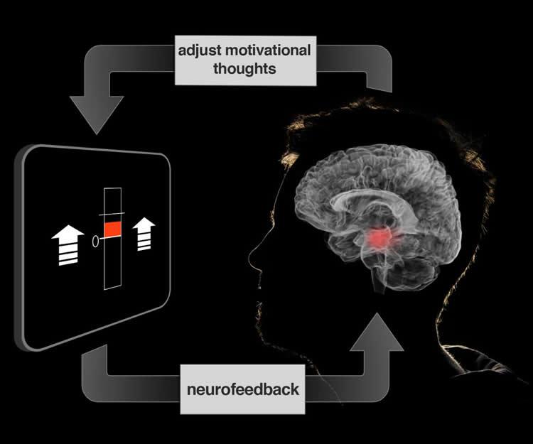
Credit: Jeff MacInnes, Duke University. Although neurofeedback has traditionally relied on the use of EEG, researchers from Duke University have examined the use of fMRI data. Participants’ brain activity was monitored and feedback was provided to them in the form of a fluctuating thermometer. Both EEG and fMRI neurofeedback were discussed in a recent article in NeuroscienceNews:
At our best, we motivate ourselves every day to get dressed and go to work or school. Although there are larger incentives at work, it’s our own volition that powers us through our innumerable daily tasks.
If we could learn to control the motivational centers of our brains that drive volition, would it lead us toward healthier, more productive lives? Using a new brain imaging strategy, Duke University scientists have now taken a first step in understanding how to manipulate specific neural circuits using thoughts and imagery.
The technique, which is described in the March 16 issue of the journal Neuron, is part of a larger approach called ‘neurofeedback,’ which gives participants a dynamic readout of brain activity, in this case from a brain area critical for motivation.
“These methods show a direct route for manipulating brain networks centrally involved in healthy brain function and daily behavior,” said the study’s senior investigator R. Alison Adcock, an assistant professor of psychiatry and behavioral sciences and associate director of the Center for Cognitive Neuroscience in the Duke University Institute for Brain Sciences.
Neurofeedback is a specialized form of biofeedback, a technique that allows people to monitor aspects of their own physiology, such as heart rate and skin temperature. It can help generate strategies to overcome anxiety and stress or to cope with other medical conditions.
Neurofeedback has historically relied on electroencephalography or EEG, in which patterns of electrical activity are monitored noninvasively by electrodes attached to the scalp. But these measures provide only rough estimates of where activity occurs in the brain.
In contrast, the new study employed functional magnetic resonance imaging (fMRI), which measures changes in blood oxygen levels, allowing more precisely localized measurements of brain activity.
Adcock’s team has been working on ways to use thoughts and behavior to tune brain function for the past eight years. In this time, they’ve developed tools allowing them to analyze complex brain imaging data in real time and to display it to participants as neurofeedback while they are in the fMRI scanner.
This study focused on the ventral tegmental area (VTA), a small area deep within the brain that is a major source of dopamine, a neurochemical well known for its role in motivation, experiencing rewards, learning, and memory.
According to Adcock’s previous research, when people are given incentives to remember specific images, an increase in VTA activation before the image appears predicts whether the participants are going to successfully remember the image.
External incentives like money work well to stimulate the VTA, but it was unclear whether people could exercise this area on their own, said co-author Jeff MacInnes, a postdoctoral researcher in Adcock’s lab.
In the new study, the team encouraged participants in the scanner to generate feelings of motivation – using their own personal strategies – during 20-second intervals. They weren’t able to raise their VTA activity consistently on their own.
But when the scientists provided participants with neurofeedback from the VTA, presented in the form of a fluctuating thermometer, participants were able to learn which strategies worked, and ultimately adopt more effective strategies. Compared to control groups, the neurofeedback-trained participants successfully elevated their VTA activity.
Participants reported using a variety of different motivational strategies, from imagining parents or coaches encouraging them, to playing out hypothetical scenarios in which their efforts were rewarded, said co-author Kathryn Dickerson, a postdoctoral researcher in Adcock’s group.
The self-generated boost in VTA activation worked even after the thermometer display was removed. Only the participants who had received accurate neurofeedback were able to consistently raise their VTA levels.
“Because this is the first demonstration of its kind, there is much still to be understood,” Adcock added. “But these tools could offer benefits for everyone, particularly those with depression or attention problems.”
The neurofeedback training also activated other regions involved in learning and experiencing rewards, confirming that, at least in the short term, the brain changes its activity more broadly as a result of neurofeedback, Dickerson said.
Adcock said one caveat of the study is that the team has not tested whether the neurofeedback drove changes in behavior. The group is working on those studies now and also plans to conduct the same study in participants with depression and attention deficit hyperactivity disorder (ADHD).
Read the original article Here
- Comments (0)
- Exercise Boosts Neurotransmitters
- By Jason von Stietz, M.A.
- March 18, 2016
-

Getty Images Why does exercise seem to improve our mood? Depression often results in the depletion of two key neurotransmitters: glutamate and GABA. However, recent findings suggest that vigorous exercise boosts the production of these neurotransmitters in the brain. Researchers at University of California Davis examined the effect of exercise on the brain using MRI scans. The study was discussed in a recent article in NeuroScientistNews:
"Major depressive disorder is often characterized by depleted glutamate and GABA, which return to normal when mental health is restored," said study lead author Richard Maddock, professor in the Department of Psychiatry and Behavioral Sciences. "Our study shows that exercise activates the metabolic pathway that replenishes these neurotransmitters."
The research also helps solve a persistent question about the brain, an energy-intensive organ that consumes a lot of fuel in the form of glucose and other carbohydrates during exercise. What does it do with that extra fuel?
"From a metabolic standpoint, vigorous exercise is the most demanding activity the brain encounters, much more intense than calculus or chess, but nobody knows what happens with all that energy," Maddock said. "Apparently, one of the things it's doing is making more neurotransmitters."
The striking change in how the brain uses fuel during exercise has largely been overlooked in brain health research. While the new findings account for a small part of the brain's energy consumption during exercise, they are an important step toward understanding the complexity of brain metabolism. The research also hints at the negative impact sedentary lifestyles might have on brain function, along with the role the brain might play in athletic endurance.
"It is not clear what causes people to 'hit the wall' or get suddenly fatigued when exercising," Maddock said. "We often think of this point in terms of muscles being depleted of oxygen and energy molecules. But part of it may be that the brain has reached its limit."
To understand how exercise affects the brain, the team studied 38 healthy volunteers. Participants exercised on a stationary bicycle, reaching around 85 percent of their predicted maximum heart rate. To measure glutamate and GABA, the researchers conducted a series of imaging studies using a powerful 3-tesla MRI to detect nuclear magnetic resonance spectra, which can identify several compounds based on the magnetic behavior of hydrogen atoms in molecules.
The researchers measured GABA and glutamate levels in two different parts of the brain immediately before and after three vigorous exercise sessions lasting between eight and 20 minutes, and made similar measurements for a control group that did not exercise. Glutamate or GABA levels increased in the participants who exercised, but not among the non-exercisers. Significant increases were found in the visual cortex, which processes visual information, and the anterior cingulate cortex, which helps regulate heart rate, some cognitive functions and emotion. While these gains trailed off over time, there was some evidence of longer-lasting effects.
"There was a correlation between the resting levels of glutamate in the brain and how much people exercised during the preceding week," Maddock said. "It's preliminary information, but it's very encouraging."
These findings point to the possibility that exercise could be used as an alternative therapy for depression. This could be especially important for patients under age 25, who sometimes have more side effects from selective serotonin reuptake inhibitors (SSRIs), anti-depressant medications that adjust neurotransmitter levels.
For follow-up studies, Maddock and the team hope to test whether a less-intense activity, such as walking, offers similar brain benefits. They would also like to use their exercise-plus-imaging method on a study of patients with depression to determine the types of exercise that offer the greatest benefit.
"We are offering another view on why regular physical activity may be important to prevent or treat depression," Maddock said. "Not every depressed person who exercises will improve, but many will. It's possible that we can help identify the patients who would most benefit from an exercise prescription."
Read the original article Here
- Comments (0)
- Studying Prejudice Using EEG
- By Jason von Stietz, M.A.
- March 5, 2016
-
Getty Images Generally, people hold more positive associations toward members of their “in-group” than of an outgroup. For example, sports fan would be quicker to bring to mind words such as “talented” or “great” when describing their favorite team than when describing other teams. This same process has been found to play out when relating to cultural or ethnic in-groups. People are quicker to bring to mind positive associations when presented with an image of someone from their own cultural background. The literature has suggested that this process is involved in unconscious prejudice in which individuals are slower to relate positive attributes to people who are different from them. Researchers from University of Bern investigated this process using EEG. The study was discussed in a recent article in NeuroscienceNews:
We do not always say what we think: we like to hide certain prejudices, sometimes even from ourselves. But unconscious prejudices become visible with tests, because we need a longer time if we must associate unpleasant things with positive terms. Researchers in Bern now show that additional processes in the brain are not responsible for this, but some of them simply take longer.
A soccer fan needs more time to associate a positive word with an opposing club than with his own team. And supporters of a political party associate a favourable attribute faster with their party than with political rivals – even if they endeavour towards the opposite. It is long since known that a positive association with one’s own group, an “in-group”, happens unconsciously faster than with an “outgroup”. These different reaction times become visible in the Implicit Association Test (IAT) with which psychologists examine unconscious processes and prejudices. But why the effort to address a friendly word to an outgroup takes more time was not clear up to now.
Now a team headed by Prof. Daria Knoch from the Department of Social Psychology and SocialNeuroscience at the Institute of Psychology, University of Bern, shows that an additional mental process is not responsible for this, as has often been postulated – but rather the brain lingers longer in certain processes. The study has now been published in the scientific journal PNAS.
Number and sequence of processes are exactly the same
The researchers relied on a unique combination of methods for their study: they conducted an Implicit Association Test with 83 test subjects who are soccer fans or political supporters. While the test persons had to associate positive terms on the screen by means of a button click, either with their in-group or with an outgroup, the brain activity was recorded by means of an EEG (electroen- cephalogram). “We analysed these data with a so-called “microstate analysis”. It enabled us to depict all processes in the brain for the first time – from the presentation of a word up to pressing the button – temporally and also spatially”, explains co-lead author Dr. Lorena Gianotti from the Department of Social Psychology and Social Neuroscience.
The analysis shows the following: the brain runs through seven processes, from the presentation of stimulus – i.e. a word – up to button click, in less than one second. “The number and sequences of these processes remain exactly the same, regardless of whether the test subject had to associate positive words with the in-group, i.e. their club or their party, or with an outgroup”, explains co-lead author Dr. Bastian Schiller, who is in the meantime conducting research at the University of Freiburg.
The reaction time with the outgroup situation is therefore longer, because some of the seven processes take longer – and not because a new process is switched in between. “As a result, corresponding theories can be refuted”, says Schiller. A complete consideration of all processes in the brain is essential for an interpretation, emphasises Lorena Gianotti, and she illustrated this in the following example: on Monday after work you go out to eat with a friend and go to sleep afterwards at 10 pm. On Friday you do exactly the same thing – but you come home two hours later since you can sleep late on the next day. If you now compare the days at 8 pm, both times you were in a restaurant and one could conclude that this is an identical time schedule. If the comparison takes place at 11 pm, you are one time already in bed and one time still on the go. One could think that on Friday you were perhaps still in the sports studio or had an entirely different daily schedule. Therefore it is clear that selective considerations do not allow any conclusion with regard to the entire day – neither with regard to the sequence nor the activities.
“In the research of human behaviour it is essential to consider the underlying brain mechanisms. And this in turn requires suitable methods in order to gain comprehensive findings”, summarises study leader Daria Knoch. A combination of neuroscientific and psychological methods can lead to new insights.
Read the original article Here
- Comments (0)
 Subscribe to our Feed via RSS