Blog
- Brain Connectivity Related Ability to Learn New Language
- By Jason von Stietz, M.A.
- January 29, 2016
-

Getty Images Why do some adults find it easier to learn a second language than others do? Recent research suggests that one’s ability to learn a new language is related to brain connectivity. Researchers from McGill University found that fMRI scans showed the greater connectivity between the left anterior insula/frontal operculum (AI/FO) and the visual word form area (VWFA) in English speakers able to learn French more quickly than their peers. The article was discussed in a recent article of NeuroScientistNews:
Learning a second language is easier for some adults than others, and innate differences in how the various parts of the brain “talk” to one another may help explain why, according to a new study published in The Journal of Neuroscience.
“These findings have implications for predicting language learning success and failure,” said study author Xiaoqian Chai.
The various regions of our brains communicate with each other even when we are resting and aren’t engaged in any specific tasks. The strength of these connections—called resting-state connectivity—varies from person to person, and differences have previously been linked to differences in behavior including language ability.
Led by Chai and Denise Klein, researchers at McGill University explored whether differences in resting-state connectivity relate to performance in a second language. To study this, the group at the Montreal Neurological Institute scanned the brains of 15 adult English speakers who were about to begin an intensive 12-week French course, and then tested their language abilities both before and after the course.
Using resting state functional magnetic resonance imaging (fMRI), the researchers examined the connectivity within the subjects’ brains prior to the start of the French course. They looked at the strength of connections between various areas in the brain and two specific language regions: an area of the brain implicated in verbal fluency, the left anterior insula/frontal operculum (AI/FO), and an area active in reading, the visual word form area (VWFA).
The researchers tested the participants’ verbal fluency and reading speed both prior to the course and after its completion. To test verbal fluency, the researchers gave subjects a prompt and asked them to speak for two minutes in French. The researchers counted the number of unique words that were used correctly. To test reading speed, the researchers had participants read French passages aloud, and they calculated the number of words read per minute.
Participants with stronger connections between the left AI/FO and an important region of the brain’s language network called the left superior temporal gyrus showed greater improvement in the speaking test. Participants with greater connectivity between the VWFA and a different area of the left superior temporal gyrus language area in the left temporal lobe showed greater improvement in reading speed by the end of the 12-week course.
“The most interesting part of this finding is that the connectivity between the different areas was observed before learning,” said Arturo Hernandez, a neuroscientist at the University of Houston who studies second-language learning and was not involved in the study. “This shows that some individuals may have a particular neuronal activity pattern that may lend itself to better learning of a second language.”
However, that doesn’t mean success at a second language is entirely predetermined by the brain’s wiring. The brain is very plastic, meaning that it can be shaped by learning and experience, Chai said.
The study is “a first step to understanding individual differences in second language learning,” she added. “In the long term it might help us to develop better methods for helping people to learn better.”
See the original article Here
- Comments (0)
- Poverty Linked to Brain Connectivity and Depression
- By Jason von Stietz, M.A.
- January 25, 2016
-

Getty Images Poverty has been linked to a litany of negative outcomes. Now, altered brain connectivity is among them. Researchers at Washington University St. Louis found that that poverty was associated with reduced connectivity between the hippocampus and the amygdala and surrounding brain regions, which in turn, was associated with higher levels of depression. The study was discussed in a recent article in MedicalXpress:
Many negative consequences are linked to growing up poor, and researchers at Washington University St. Louis have identified one more: altered brain connectivity.
Analyzing brain scans of 105 children ages 7 to 12, the researchers found that key structures in the brain are connected differently in poor children than in kids raised in more affluent settings. In particular, the brain's hippocampus—a structure key to learning, memory and regulation of stress—and the amygdala—which is linked to stress and emotion—connect to other areas of the brain differently in poor children than in kids whose families had higher incomes.
Those connections, viewed using functional MRI scans, were weaker, depending on the degree of poverty to which a child was exposed. The poorer the family, the more likely the hippocampus and amygdala would connect to other brain structures in ways the researchers characterized as weaker. In addition, poorer preschoolers were much more likely to have symptoms of clinical depression when they reached school age.
The study is available online Friday, Jan. 15, in The American Journal of Psychiatry.
"Our past research has shown that the brain's anatomy can look different in poor children, with the size of the hippocampus and amygdala frequently altered in kids raised in poverty," said first author Deanna M. Barch, PhD, chair of Washington University's Department of Psychological & Brain Sciences in Arts & Sciences, and the Gregory B. Couch Professor of Psychiatry at the School of Medicine. "In this study, we found that the way those structures connect with the rest of the brain changes in ways we would consider to be less helpful in regulating emotion and stress."
Those changes in connectivity also are related to a risk of clinical depression. Those in the study who were poor as preschoolers were more likely to be depressed at age 9 or 10.
Previous research from the same group of investigators had identified differences in the volume of gray matter and white matter, and the size and volume of the hippocampus and amygdala. But they also found that many of those changes could be overcome through nurturing from parents. That wasn't true, however, regarding changes in connectivity identified in the new study.
"Poverty is one of the most powerful predictors of poor developmental outcomes for children," said co-investigator Joan L. Luby, MD, the Samuel and Mae S. Ludwig Professor of Child Psychiatry and director of Washington University's Early Emotional Development Program. "Previously, we've seen that there may be ways to overcome some brain changes linked to poverty, but we didn't see anything that reversed the negative changes in connectivity present in poor kids."
The researchers measured poverty using what's called an income-to-needs ratio that takes into account a family's size and annual income. The current federal poverty level is $24,250 for a family of four.
Children raised in poverty tend to have poorer cognitive and educational outcomes and are at higher risk for psychiatric illnesses, including depression and antisocial behaviors. Researchers hypothesize that factors such as stress, adverse environmental exposures—including lead, cigarette smoke and poor nutrition—along with limited educational opportunities, can contribute to problems later in life.
But Barch emphasized that the link between poverty and poor outcomes doesn't necessarily lock a child into a difficult life.
"Many things can be done to foster brain development and positive emotional development," she said. "Poverty doesn't put a child on a predetermined trajectory, but it behooves us to remember that adverse experiences early in life are influencing the development and function of the brain. And if we hope to intervene, we need to do it early so that we can help shift children onto the best possible developmental trajectories."
Read the original article Here
- Comments (0)
- Stories Involving Personal Values Activate Default Mode Network
- By Jason von Stietz
- January 21, 2016
-

Getty Images Stories involving infidelity, politics, war, labor strikes, or abortion often tap into our most deeply held values. Recent research found that value-laden stories activate the default mode network, previously thought to activate as the brain’s autopilot. Researchers now believe the default mode network is activated as the brain works to find meaning in narrative. The study was discussed in a recent article in MedicalXpress:
Everyone has at least a few non-negotiable values. These are the things that, no matter what the circumstance, you'd never compromise for any reason - such as "I'd never hurt a child," or "I'm against the death penalty."
Real-time brain scans show that when people read stories that deal with these core, protected values, the "default mode network" in their brains activates.
This network was once thought of as just the brain's autopilot, since it has been shown to be active when you're not engaged by anything in the outside world - but studies like this one suggest that it's actually working to find meaning in the narratives.
"The brain is devoting a huge amount of energy to whatever that network is doing. We need to understand why," said Jonas Kaplan of the USC Dornsife Brain and Creativity Institute. Kaplan was the lead author of the study, which was published on Jan. 7 in the journal Cerebral Cortex.
Kaplan thinks that it's not just that the brain is presented with a moral quandary, but rather that the quandary is presented in a narrative format.
"Stories help us to organize information in a unique way," he said.
To find relevant stories, the researchers sorted through 20 million blog posts using software developed at the USC Institute for Creative Technologies.
"We wanted to know how people tell stories in their daily lives. It was kind of like finding stories in their natural habitat," said Kaplan, assistant research professor of psychology at the Brain and Creativity Institute at the USC Dornsife College of Letters, Arts and Sciences.
That 20 million was pared down to 40 stories that each contained an example of a crisis involving a potentially protected value: cheating on a spouse, having an abortion, crossing a picket line, or getting in a fight.
Those stories were translated into Mandarin Chinese and Farsi, and then read by American, Chinese and Iranian participants in their native language while their brains were scanned by fMRI. They also answered general questions about the stories while being scanned.
Stories that participants said involved values that were protected to them activated the default mode network in their brain to a greater degree. In addition, the level of activation varied from culture to culture. On average, Iranians showed the greatest level of activation in the study, while the Chinese participants showed the least.
"Stories appear to be a fundamental way in which the brain organizes information in a practical and memorable manner. It is important to understand the neural mechanisms required to do this, and this study is a step in that direction," said Antonio Damasio, senior author of the study. Damasio is co-director of the Brain and Creativity Institute, holder of the David Dornsife Chair in Neuroscience and a professor of psychology and neurology.
It's not yet clear whether a value either is or is not protected, or whether the sacredness of a value is on a sliding scale. But in a nation where political beliefs are growing more polarized and entrenched, it's important to understand what biological processes lie at the root of these values, Kaplan said.
"People will often hold political values as protected values and protected values are at the root of many political conflicts around the world, which is why they're interesting to us," he said.
Read the original article Here
- Comments (0)
- Brainwaves Spread Through Mild Electrical Field
- By Jason von Stietz, M.A.
- January 15, 2016
-
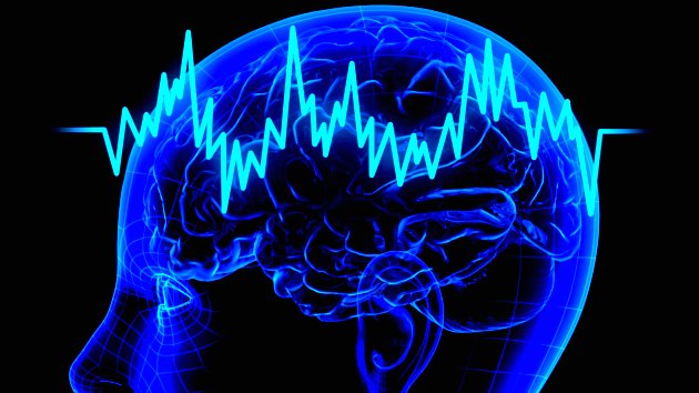
iStockPhoto Scientists once thought that the brain’s electrical endogenous fields were too weak to propagate transmission. However, recent findings from researchers from Case Western Reserve University suggested that brainwaves spread through a mild electrical field. The study was discussed in a recent article in MedicalXpress:
Researchers at Case Western Reserve University may have found a new way information is communicated throughout the brain.
Their discovery could lead to identifying possible new targets to investigate brain waves associated with memory and epilepsy and better understand healthy physiology.
They recorded neural spikes traveling at a speed too slow for known mechanisms to circulate throughout the brain. The only explanation, the scientists say, is the wave is spread by a mild electrical field they could detect. Computer modeling and in-vitro testing support their theory.
"Others have been working on such phenomena for decades, but no one has ever made these connections," said Steven J. Schiff, director of the Center for Neural Engineering at Penn State University, who was not involved in the study. "The implications are that such directed fields can be used to modulate both pathological activities, such as seizures, and to interact with cognitive rhythms that help regulate a variety of processes in the brain."
Scientists Dominique Durand, Elmer Lincoln Lindseth Professor in Biomedical Engineering at Case School of Engineering and leader of the research, former graduate student Chen Sui and current PhD students Rajat Shivacharan and Mingming Zhang, report their findings in The Journal of Neuroscience.
"Researchers have thought that the brain's endogenous electrical fields are too weak to propagate wave transmission," Durand said. "But it appears the brain may be using the fields to communicate without synaptic transmissions, gap junctions or diffusion."
How the fields may work
Computer modeling and testing on mouse hippocampi (the central part of the brain associated with memory and spatial navigation) in the lab indicate the field begins in one cell or group of cells.
Although the electrical field is of low amplitude, the field excites and activates immediate neighbors, which, in turn, excite and activate immediate neighbors, and so on across the brain at a rate of about 0.1 meter per second.
Blocking the endogenous electrical field in the mouse hippocampus and increasing the distance between cells in the computer model and in-vitro both slowed the speed of the wave.
These results, the researchers say, confirm that the propagation mechanism for the activity is consistent with the electrical field.
Because sleep waves and theta waves—which are associated with forming memories during sleep—and epileptic seizure waves travel at about 1 meter per second, the researchers are now investigating whether the electrical fields play a role in normal physiology and in epilepsy.
If so, they will try to discern what information the fields may be carrying. Durand's lab is also investigating where the endogenous spikes come from.
Read the original article Here
- Comments (1)
 Subscribe to our Feed via RSS