Blog
- Autism Biomarker for Boys Found
- By Jason von Stietz, M.A.
- April 29, 2016
-
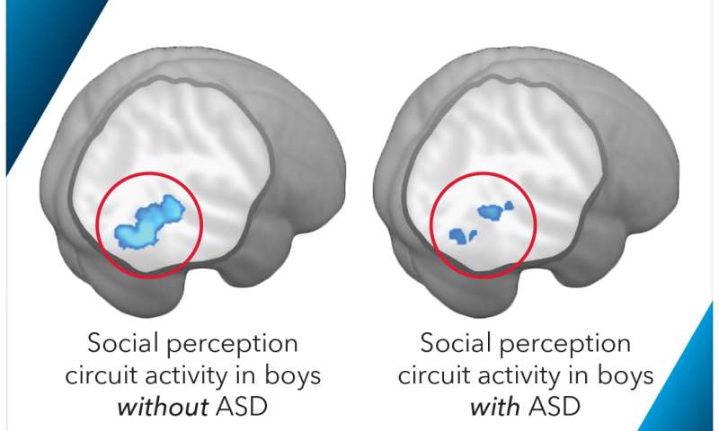
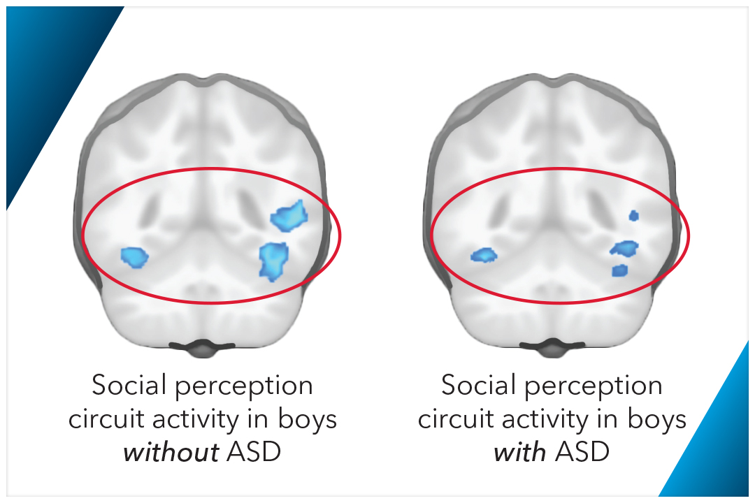
Credit: George Washington University Researchers at the George Washington University investigated the use of brain scans in measuring the progress of treatments for Autism Spectrum Disorder in boys. Findings indicated that a biomarker related to brain circuitry involved in social perception may assist in more quickly diagnosing and treating difficult to diagnose patients. The study was discussed in a recent article in Medical Xpress:
Researchers have developed a new method to map and track the function of brain circuits affected by autism spectrum disorder (ASD) in boys using brain imaging. The technique will provide clinicians and therapists with a physical measure of the progress patients are making with behavioral and/or drug treatments - a tool that has been elusive in autism treatment until this point.
For the first time, doctors would be able to quantify how that brain circuit is working in their patients and assess the effectiveness of an intervention. The research is outlined in a paper, "Quantified Social Perception Circuit Activity as a Neurobiological Marker of Autism Spectrum Disorder," published Wednesday in JAMA Psychiatry. The paper focuses on the use of biomarkers, measurable indicators of a biological condition, to measure the function of the social perception circuit of the brain.
"This is significant because biomarkers give us a 'why' for understanding autism in boys that we haven't had before," said Kevin Pelphrey, a co-author of the paper, who is the Carbonell Family Professor in Autism and Neurodevelopmental Disorders and director of the Autism and Neurodevelopmental Disorders Institute at the George Washington University. "We can now use functional biomarkers to identify what treatments will be effective for individual cases and measure progress."
Researchers analyzed a series of 164 images from each of 114 individuals and discovered the brain scans of the social perception circuits only indicated ASD in boys. This new research has the potential to improve treatment for ASD by measuring changes in the social perception brain circuit in response to different interventions. The researchers found the brain scan data can be an effective indicator of function of the circuit in younger children and older patients alike.The research is particularly relevant for ASD patients who are difficult to diagnose and treat by providing a more definitive diagnosis and in developing a treatment program when it is not clear if behavioral, drug or a combination of the treatments will be most effective.
"The behavioral symptoms of ASD are so complex and varied it is difficult to determine whether a new treatment is effective, especially within a realistic time frame," said Malin Björnsdotter, assistant professor at the University of Gothenburg and lead author of the paper. "Brain function markers may provide the specific and objective measures required to bridge this gap."
A Path to Widespread use of Brain Scans?
In addition to helping to identify the most effective ASD treatment for an individual, this research provides evidence that brain imaging is an important intervention tool. Currently, functional MRI, the type of brain scan used in this study, is not a standard part of ASD treatment, as there is not enough evidence linking the scan to effective treatments. The Autism and Neurodevelopmental Disorders Institute at GW aims to make significant contributions toward the establishment of evidence-based therapies for ASD.
Credit: George Washington University "This kind of imaging can help us answer the question, 'On day one of treatment, will this child benefit from a 16-week behavioral intervention?'" Dr. Pelphrey said. "Answering that question will help parents save time and money on diagnosis and treatments."
Following the study, Dr. Pelphrey and his colleagues will test their findings at the next level: studying a larger pool of people with autism and other neurological disorders in collaboration with Children's National Medical Center to see if the scan can successfully distinguish ASD from other disorders and track treatment progress.
The authors emphasized that this research is still in the earliest days, pointing out that doctors' offices and most hospitals do not have the specialized imaging equipment necessary to carry out the brain scans used by the team involved in this study.
"To really help patients we need to develop inexpensive, easy-to-use techniques that can be applied in any group, including infants and individuals with severe behavioral problems," said Dr. Björnsdotter. "This study is a first step toward that goal."
While this method currently only works for boys with autism, the researchers are leading a large-scale, nationwide study of girls with autism to identify equivalent techniques that will work for them. The group expects to have the initial results from that study later this year.
Read the original article Here
- Comments (0)
- Anticholinergics Linked to Changes in Brain and Cognitive Impairment
- By Jason von Stietz, M.A.
- April 26, 2016
-

Getty Images Are over the counter drugs completely safe? Researchers at Indiana University School of Medicine used positron emission tests (PET) to study the relationship between anticholinergic drugs and cognitive decline. Anticholinergics are commonly used as sleep aids as wells as treatments for hypertension and cardiovascular disease. Findings linked the use of anticholinergics to physical changes in the brain and cognitive impairment. The study was discussed in a recent article in Medical Xpress:
Older adults might want to avoid a using class of drugs commonly used in over-the-counter products such as nighttime cold medicines due to their links to cognitive impairment, a research team led by scientists at Indiana University School of Medicine has recommended.
Using brain imaging techniques, the researchers found lower metabolism and reduced brain sizes among study participants taking the drugs known to have an anticholinergic effect, meaning they block acetylcholine, a nervous system neurotransmitter.
Previous research found a link between between the anticholinergic drugs and cognitive impairment and increased risk of dementia. The new paper published in the journal JAMA Neurology, is believed to be the first to study the potential underlying biology of those clinical links using neuroimaging measurements of brain metabolism and atrophy.
"These findings provide us with a much better understanding of how this class of drugs may act upon the brain in ways that might raise the risk of cognitive impairment and dementia," said Shannon Risacher, Ph.D., assistant professor of radiology and imaging sciences, first author of the paper, "Association Between Anticholinergic Medication Use and Cognition, Brain Metabolism, and Brain Atrophy in Cognitively Normal Older Adults."
"Given all the research evidence, physicians might want to consider alternatives to anticholinergic medications if available when working with their older patients," Dr. Risacher said.
Drugs with anticholinergic effects are sold over the counter and by prescription as sleep aids and for many chronic diseases including hypertension, cardiovascular disease, and chronic obstructive pulmonary disease.
A list of anticholinergic drugs and their potential impact is athttp://www.agingbraincare.org/uploads/products/ACB_scale_-_legal_size.pdf.
Scientists have linked anticholinergic drugs cognitive problems among older adults for at least 10 years. A 2013 study by scientists at the IU Center for Aging Research and the Regenstrief Institute found that drugs with a strong anticholinergic effect cause cognitive problems when taken continuously for as few as 60 days. Drugs with a weaker effect could cause impairment within 90 days.
The current research project involved 451 participants, 60 of whom were taking at least one medication with medium or high anticholinergic activity. The participants were drawn from a national Alzheimer's research project—the Alzheimer's Disease Neuroimaging Initiative—and the Indiana Memory and Aging Study.
To identify possible physical and physiological changes that could be associated with the reported effects, researchers assessed the results of memory and other cognitive tests, positron emission tests (PET) measuring brain metabolism, and magnetic resonance imaging (MRI) scans for brain structure.
The cognitive tests revealed that patients taking anticholinergic drugs performed worse than older adults not taking the drugs on short-term memory and some tests of executive function, which cover a range of activities such as verbal reasoning, planning, and problem solving.
Anticholinergic drug users also showed lower levels of glucose metabolism—a biomarker for brain activity—in both the overall brain and in the hippocampus, a region of the brain associated with memory and which has been identified as affected early by Alzheimer's disease.
The researchers also found significant links between brain structure revealed by the MRI scans and anticholinergic drug use, with the participants using anticholinergic drugs having reduced brain volume and larger ventricles, the cavities inside the brain.
"These findings might give us clues to the biological basis for the cognitive problems associated with anticholinergic drugs, but additional studies are needed if we are to truly understand the mechanisms involved," Dr. Risacher said.
Read the original article Here
- Comments (0)
- Study Links Affective Understanding to Attraction
- By Jason von Stietz, M.A.
- April 8, 2016
-

Getty Images What is it that leads to attraction? Researchers conducted an experiment utilizing fMRI scans of participants watching a video of a woman. It was found that participants found her more attractive, which was reflected in the reward center of the brain, when they perceived themselves to understand her emotional experience. The findings were discussed in a recent article in Medical Xpress:
(Medical Xpress)—A team of researchers with members from a large number of institutions in Germany has conducted a study that has revealed more about the way interpersonal attraction works in the brain. In their paper published in Proceedings of the National Academy of Sciences, the group describes two experiments they conducted with volunteers, their results and what they believe was revealed about the nature of the mechanism of attraction between people.
Most everyone has experienced near instant attraction to someone else, whether of a social or sexual nature, but few are able to pin down exactly why they felt that attraction. Based on two experiments they conducted with human volunteers, the researchers suggest it may have to do with matchingneural circuitry.
To learn more about attraction, the researchers ran two experiments, the first consisted of showing 19 male and 21 female volunteers, videos of six different women as they experienced fear or sadness. The volunteers were asked to choose which emotion was being shown, and then to mark down how confident they were in their choice. To gauge how much of an attraction they volunteers felt for the women in the videos, they were asked to enlarge a picture of the woman both before and after seeing her in the video—each was also asked to answer questions about each woman, such as how much they would like to meet her in real life, if she would understand them, etc.
The second experiment was run with a different set of volunteers who were also asked to watch the woman in the videos, but the second group did so while undergoing an fMRI imaging—the researchers were specifically looking for activity in the part of the brain known to be associated with rewards.
The final phase of the experiment involved combining data from both experiments to see if any patterns might emerge. The researchers report that most of the volunteers were able to identify the emotions being portrayed, and the more confident they felt they were able to identify the correct emotion, the more attracted to her they felt. This was confirmed in the fMRI scans—reward centers in the volunteers' brains lit up more when watching women they felt they could read their emotions better.
The researchers propose that their results suggest that in addition to physical attractiveness, people are attracted to other people due to their own feelings of similarity to another person, which gives them a feeling of understanding, or connectedness.
Read the original article Here
- Comments (0)
- Neuronal Feedback Changes Our Perception
- By Jason von Stietz, M.A.
- April 4, 2016
-

Photo Credit: Carnegie Mellon University Have you ever thought you saw one thing, but when you looked again it was something else? Often times the brain notices the beginning of a visual pattern and will automatically connect the rest of the dots. How does this happen? Researchers at the Carnegie Mellon University studied top-down visual processing. Findings were discussed in a recent article in MedicalXpress:
Understanding this feedback system could provide new insight into thevisual system's neuronal circuitry and could have further implications for understanding how the brain interprets and understands sensory stimuli.
Many optical illusions make you see something that's not there. Take the Kanizsa triangle: when you place three Pac-Man-like wedges in the right spot, you see a triangle, even though the edges of the triangle aren't drawn.
"We see with both our brain and our eyes. Your brain is making inferences that allow you to see the triangle. It's connecting the dots between the corners of the wedges," said Kuhlman, who is a member of Carnegie Mellon's BrainHub neuroscience initiative and the joint Carnegie Mellon/University of Pittsburgh Center for the Neural Basis of Cognition (CNBC). "Optical illusions illustrate some of the amazing things our visual system can do."
When we look at an object, information about what we see travels through circuits of neurons beginning in the retina, through the thalamus and into the brain's visual cortex. In the visual cortex, the information gets processed in multiple stages and is ultimately sent to the prefrontal cortex—the area of the brain that makes decisions, including how to respond to a given stimulus.
However, not all information stays on this forward moving path. At the secondary stage of processing in the visual cortex some neurons reverse course and send information back to the first stage of processing. Researchers at Carnegie Mellon wondered if this feedback could change how the neurons in the visual cortex respond to a stimulus and alter the messages being sent to the prefrontal cortex.
While there has been a good deal of research studying how information moves forward through the visual system, less has been done to study the impact of the information that moves backward. To find out if the information traveling from the secondary stage of processing back to the first stage impacted how information is encoded in the visual system, the researchers needed to quantify the magnitude of information that was being sent from the second stage back to the first stage. Using a mouse model, they recorded normal neuronal firing in the first stage of the visual cortex as the mouse looked at moving patterns that represented edges. They then silenced the neurons in the second stage using modified optogenetic technology. This halted the feedback of information from the second stage back to the first stage, and allowed the researchers to determine how much of the neuronal activity in the first stage of visual processing was the result of feedback.
Twenty percent of the neuronal activity in the visual cortex was the result of feedback, a concept Kuhlman calls reciprocal connectivity. This indicates that some of the information coming from the visual cortex is not a direct response to a visual stimuli, but is a response to how the stimuli was perceived by higher cortical areas.
The feedback, she says, might be what causes our brain to complete the undrawn lines in the Kanizsa triangle. But more importantly, it signifies that studying neuronal feedback is important to our understanding of how the brain works to process stimuli.
"This represents a new way to study visual perception and neural computation. If we want to truly understand the visual pathway, and cortical function in general, we have to understand these reciprocal connection," Kuhlman said.
Read the original artice Here
- Comments (0)
 Subscribe to our Feed via RSS