Blog
- Brain Implant Boosts Short-Term and Working Memory
- By Jason von Stietz, MA
- November 30, 2017
-
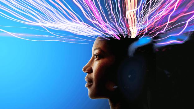
Researchers from University of Southern California have used a brain implant to help the brain’s of study participants function more effectively. Electrodes were implanted into the brain’s of participants. Once the pattern of activity associated with optimal memory performance was determined, the electrode was used to stimulate the brain reinforcing the optimal pattern. Findings indicated the short-term memory was improved by 15% and working memory was improved by 25%. The study was discussed in a recent article in Futurism:
With everyone from Elon Musk to MIT to the U.S. Department of Defense researching brain implants, it seems only a matter of time before such devices are ready to help humans extend their natural capabilities. Now, a professor from the University of Southern California (USC) has demonstrated the use of a brain implant to improve the human memory, and the device could have major implications for the treatment of one of the U.S.’s deadliest diseases.
Dong Song is a research associate professor of biomedical engineering at USC, and he recently presented his findings on a “memory prosthesis” during a meeting of the Society for Neuroscience in Washington D.C. According to a New Scientist report, the device is the first to effectively improve the human memory.
To test his device, Song’s team enlisted the help of 20 volunteers who were having brain electrodes implanted for the treatment of epilepsy.
Once implanted in the volunteers, Song’s device could collect data on their brain activity during tests designed to stimulate either short-term memory or working memory. The researchers then determined the pattern associated with optimal memory performance and used the device’s electrodes to stimulate the brain following that pattern during later tests.
Based on their research, such stimulation improved short-term memory by roughly 15 percent and working memory by about 25 percent. When the researchers stimulated the brain randomly, performance worsened.
As Song told New Scientist, “We are writing the neural code to enhance memory function. This has never been done before.”
A GROWING PROBLEM
While a better memory could be useful for students cramming for tests or those of us with trouble remembering names, it could be absolutely life-changing for people affected by dementia and Alzheimer’s.
As Bill Gates noted when announcing plans to invest $100 million of his own money into dementia and Alzheimer’s research, the disease is a multi-level problem that’s positioned to get even worse.
Age is the greatest risk factor for Alzheimer’s, according to the Alzheimer’s Association, with the vast majority of sufferers over the age of 65. With advances in medicine and healthcare continuously increasing how long we live, that segment of the population is growing dramatically, and by 2030, 20 percent of U.S. citizens are expected to be older than 65.
This increase in the number of potential dementia sufferers can be costly in both a financial and emotional sense. In 2016, the total cost of healthcare and long-term care for those suffering from dementia and Alzheimer’s disease was an estimated $236 billion, and according to the Alzheimer’s Association, the more severe a person’s cognitive impairment, the higher the rates of depression in their familial caregivers.
Of course, further testing is required before Song’s device could be approved as a treatment for dementia or Alzheimer’s, but if it is able to help those patients regain even part of their lost memory function, the impact would be felt not only by the patients themselves, but their families and even the economy at large.
Read the article Here
- Comments (0)
- Neural Signatures of Suicidal Thoughts Detected by fMRI
- By Jason von Stietz, MA
- November 26, 2017
-
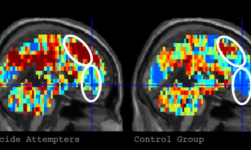
Credit: Carnegie Mellon University Can suicidal thoughts be detected by a machine? Researchers from Carnegie Mellon University among other institutions investigated the use of fMRI brain scans and machine-learning, the ability of a machine to learn without it being explicitly programmed, in the detection of neural signals related to suicidal thoughts. The researchers were able to identify individuals who had attempted suicide with 94% accuracy. The study was discussed in a recent article in MedicalXpress:
Suicidal risk is notoriously difficult to assess and predict, and suicide is the second-leading cause of death among young adults in the United States. Published in Nature Human Behaviour, the study offers a new approach to assessing psychiatric disorders.
"Our latest work is unique insofar as it identifies concept alterations that are associated with suicidal ideation and behavior, using machine-learning algorithms to assess the neural representation of specific concepts related to suicide. This gives us a window into the brain and mind, shedding light on how suicidal individuals think about suicide and emotion related concepts. What is central to this new study is that we can tell whether someone is considering suicide by the way that they are thinking about the death-related topics," said Just, the D.O. Hebb University Professor of Psychology in CMU's Dietrich College of Humanities and Social Sciences.
For the study, Just and Brent, who holds an endowed chair in suicide studies and is a professor of psychiatry, pediatrics, epidemiology and clinical and translational science at Pitt, presented a list of 10 death-related words, 10 words relating to positive concepts (e.g. carefree) and 10 words related to negative ideas (e.g. trouble) to two groups of 17 people with known suicidal tendencies and 17 neurotypical individuals.
They applied the machine-learning algorithm to six word-concepts that best discriminated between the two groups as the participants thought about each one while in the brain scanner. These were death, cruelty, trouble, carefree, good and praise. Based on the brain representations of these six concepts, their program was able to identify with 91 percent accuracy whether a participant was from the control or suicidal group.
Then, focusing on the suicidal ideators, they used a similar approach to see if the algorithm could identify participants who had made a previous suicide attempt from those who only thought about it. The program was able to accurately distinguish the nine who had attempted to take their lives with 94 percent accuracy.
"Further testing of this approach in a larger sample will determine its generality and its ability to predict future suicidal behavior, and could give clinicians in the future a way to identify, monitor and perhaps intervene with the altered and often distorted thinking that so often characterizes seriously suicidal individuals," said Brent.
To further understand what caused the suicidal and non-suicidal participants to have different brain activation patterns for specific thoughts, Just and Brent used an archive of neural signatures for emotions (particularly sadness, shame, anger and pride) to measure the amount of each emotion that was evoked in a participant's brain by each of the six discriminating concepts. The machine-learning program was able to accurately predict which group the participant belonged to with 85 percent accuracy based on the differences in the emotion signatures of the concepts.
"The benefit of this latter approach, sometimes called explainable artificial intelligence, is more revealing of what discriminates the two groups, namely the types of emotions that the discriminating words evoke," Just said. "People with suicidal thoughts experience different emotions when they think about some of the test concepts. For example, the concept of 'death' evoked more shame and more sadness in the group that thought about suicide. This extra bit of understanding may suggest an avenue to treatment that attempts to change the emotional response to certain concepts."
Just and Brent are hopeful that the findings from this basic cognitive neuroscience research can be used to save lives.
"The most immediate need is to apply these findings to a much larger sample and then use it to predict future suicide attempts," said Brent.
Just and his CMU colleague Tom Mitchell first pioneered this application of machine learning to brain imaging that identifies concepts from their brain activation signatures. Since then, the research has been extended to identify emotions and multi-concept thoughts from their neural signatures and also to uncover how complex scientific concepts are coded as they are being learned.
Read the article Here
- Comments (0)
- Chronic Alcohol Consumption Kills Brain Stem Cells
- By Jason von Stietz, MA
- November 19, 2017
-

Chronic alcohol abuse gone untreated can lead to severe brain damage. Researchers at The University of Texas Medical Branch at Galveston recently investigated the impact of alcohol consumption on stem cells in the brains of mice. Findings indicated that chronic alcohol consumption killed brain stem cells, reduced the production of new nerve cells, and affected females significantly worse than males. The study was discussed in a recent article in MedicalXpress:
Because the brain stems cells create new nerve cells and are important to maintaining normal cognitive function, this study possibly opens a door to combating chronic alcoholism.
The researchers also found that brain stem cells in key brain regions of adult mice respond differently to alcohol exposure, and they show for the first time that these changes are different for females and males. The findings are available in Stem Cell Reports.
Chronic alcohol abuse can cause severe brain damage and neurodegeneration. Scientists once believed that the number of nerve cells in the adult brain was fixed early in life and the best way to treat alcohol-induced brain damage was to protect the remaining nerve cells.
"The discovery that the adult brain produces stem cells that create new nerve cells provides a new way of approaching the problem of alcohol-related changes in the brain," said Dr. Ping Wu, UTMB professor in the department of neuroscience and cell biology. "However, before the new approaches can be developed, we need to understand how alcohol impacts the brain stem cells at different stages in their growth, in different brain regions and in the brains of both males and females."
In the study, Wu and her colleagues used a cutting-edge technique that allows them to tag brain stem cells and observe how they migrate and develop into specialized nerve cells over time to study the impact of long-term alcohol consumption on them.
Wu said that chronic alcohol drinking killed most brain stem cells and reduced the production and development of new nerve cells.
The researchers found that the effects of repeated alcohol consumption differed across brain regions. The brain region most susceptible to the effects of alcohol was one of two brain regions where new brain cells are created in adults.
They also noted that female mice showed more severe deficits than males. The females displayed more severe intoxication behaviors and more greatly reduced the pool of stem cells in the subventricular zone.
Using this model, scientists expect to learn more about how alcohol interacts with brain stem cells, which will ultimately lead to a clearer understanding of how best to treat and cure alcoholism.
Read the article Here
- Comments (0)
 Subscribe to our Feed via RSS