Blog
- Robotic Exoskeleton Improves Walking Ability in Children With Cerebral Palsy
- By Jason von Stietz, M.A.
- August 27, 2017
-
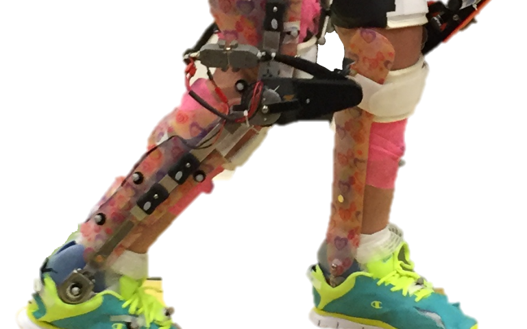
Credit: Northern Arizona University Children with cerebral palsy, a neurological and movement disorder, often limits mobility and independent life functioning in children. Researchers at Northern Arizona University and the National Institutes of Health examined the use of a robotic exoskeleton to improve waling ability in children with cerebral palsy. The study was discussed in a recent article in Medical Xpress:
According to the Centers for Disease Control and Prevention, cerebral palsy (CP)—caused by neurological damage before, during or after birth—is the most common movement disorder in children, limiting mobility and independence throughout their lives. An estimated 500,000 children in the U.S. have CP.
Although nearly 60 percent of children with the disorder can walk independently, many have crouch gait, a pathological walking pattern characterized by excessive knee bending, which can cause an abnormally high level of stress on the knee. Crouch gait can lead to knee pain and progressive loss of function and is often treated through invasive orthopedic surgery.
Assistant professor of mechanical engineering Zach Lerner, who joined Northern Arizona University's Center for Bioengineering Innovation in 2017, recently published a study in the journal Science Translational Medicine investigating whether wearing a robotic exoskeleton—a leg brace powered by small motors—could alleviate crouch gait in children with cerebral palsy.
"We evaluated a novel exoskeleton for the treatment of crouch gait, one of the most debilitating pathologies in CP," Lerner said. "In our exploratory, multi-week trial, we fitted seven participants between the ages of five and 19 with robotic exoskeletons designed to increase their ability to extend their knees at specific phases in the walking cycle."
After being fitted with the assistive devices, the children participated in several practice sessions. At the end of the trial, six of the seven participants exhibited improvements in walking posture equivalent to outcomes reported from invasive orthopedic surgery. The researchers also demonstrated that improvements in crouch increased over the course of the exploratory trial, which was conducted at the National Institutes of Health Clinical Center in Bethesda, Maryland.
"Together, these results provide evidence supporting the use of wearable exoskeletons as a treatment strategy to improve walking in children with CP," Lerner said.
The exoskeleton was safe and well-tolerated, and all the children were able to walk independently with the device. Rather than guiding the lower limbs, the exoskeleton dynamically changed their posture by introducing bursts of knee extension assistance during discrete portions of the walking cycle, which resulted in maintained or increased knee extensor muscle activity during exoskeleton use.
"Our results suggest powered knee exoskeletons should be investigated as an alternative to or in conjunction with existing treatments for crouch gait, including orthopedic surgery, muscle injections and physical therapy," Lerner said.
Lerner leads NAU's Biomechatronics Lab, where his goal is to improve mobility and function in individuals with neuromuscular and musculoskeletal disabilities through innovations in mechanical and biomedical engineering. Building on the encouraging results of this study, his team is working toward conducting longer-term exoskeleton interventions to take place at home and in the community.
View the article Here
- Comments (0)
- Fear-Memories Can Be Erased, Study Finds
- By Jason von Stietz, M.A.
- August 25, 2017
-
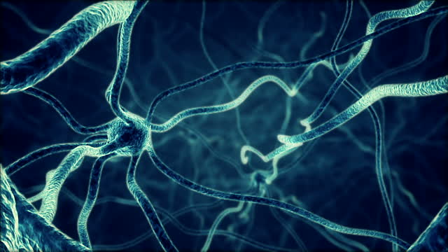
Getty Images At times, fear is healthy and necessary. For example, an individual hiking in the woods should feel fear and avoid approaching a bear or other wild animals. However, often times individuals develop unhealthy or unhelpful fears such as a phobia of dogs, even if they are domesticated and friendly. Researchers at University of California, Riverside investigatesd the use of a method of selectively erasing fear-memories by weakening the connections of the neurons involved in the formation of those memories. The study was discussed in a recent article in Medical Xpress:
To survive in a dynamic environment, animals develop fear responses to dangerous situations. But not all fear memories, such as those in PTSD, are beneficial to our survival. For example, while an extremely fearful response to the sight of a helicopter is not a useful one for a war veteran, a quick reaction to the sound of a gunshot is still desirable. For survivors of car accidents, it would not be beneficial for them to relive the trauma each time they sit in a car.
In their lab experiments, Jun-Hyeong Cho, M.D., Ph.D., an assistant professor of molecular, cell, and systems biology, and Woong Bin Kim, his postdoctoral researcher, found that fear memory can be manipulated in such a way that some beneficial memories are retained while others, detrimental to our daily life, are suppressed.
The research, done using a mouse model and published today in Neuron, offers insights into how PTSD and specific phobias may be better treated.
"In the brain, neurons communicate with each other through synaptic connections, in which signals from one neuron are transmitted to another neuron by means of neurotransmitters," said Cho, who led the research. "We demonstrated that the formation of fear memory associated with a specific auditory cue involves selective strengthening in synaptic connections which convey the auditory signals to the amygdala, a brain area essential for fear learning and memory. We also demonstrated that selective weakening of the connections erased fear memory for the auditory cue."
In the lab, Cho and Kim exposed mice to two sounds: a high-pitch tone and a low-pitch tone. Neither tone produced a fear response in the mice. Next, they paired only the high-pitched tone with a mild footshock administered to the mice. Following this, Cho and Kim again exposed the mice to the two tones. To the high-pitch tone (with no accompanying footshock), the mice responded by ceasing all movement, called freezing behavior. The mice showed no such response to the low-pitch sound (with no accompanying footshock). The researchers found that such behavioral training strengthened synaptic connections that relay the high-pitch tone signals to the amygdala.
The researchers then used a method called optogenetics to weaken the synaptic connection with light, which erased the fear memory for the high-pitch tone.
"In the brain, neurons receiving the high- and low-pitch tone signals are intermingled," said Cho, a member of the Center for Glial-Neuronal Interactions in the UC Riverside School of Medicine. "We were able, however, to experimentally stimulate just those neurons that responded to the high-pitch sound. Using low-frequency stimulations with light, we were able to erase the fear memory by artificially weakening the connections conveying the signals of the sensory cue—a high-pitch tone in our experiments - that are associated with the aversive event, namely, the footshock."
Cho explained that for adaptive fear responses to be developed, the brain must discriminate between different sensory cues and associate only relevant stimuli with aversive events.
"This study expands our understanding of how adaptive fear memory for a relevant stimulus is encoded in the brain," he said. "It is also applicable to developing a novel intervention to selectively suppress pathological fear while preserving adaptive fear in PTSD."
The researchers note that their method can be adapted for other research, such as "reward learning" where stimulus is paired with reward. They plan next to study the mechanisms involved in reward learning which has implications in treating addictive behaviors.
Read the article Here
- Comments (1)
 Subscribe to our Feed via RSS