Blog
- Is Mindfulness Different Than Relaxation?
- By Jason von Stietz, M.A.
- June 21, 2018
-

Are the effects of mindfulness meditation the same as relaxation training? Mindfulness meditation often leads to feelings of calmness. However, the goal of mindfulness is to practice non-judgmental present moment awareness and not necessarily. Does this difference in goals lead to changes in the brain’s response? Researchers at Massachusetts General Hospital led a study utilizing functional magnetic resonance imaging to examine the difference in neural activity of participants engaging in mindfulness based stress reduction versus relaxation response training. The study was discussed in a recent article in MedicalXpress:
There are two widely used meditation-based stress reduction courses. One is based on the relaxation response—first described by Herb Benson, MD, director emeritus of the MGH-based Benson-Henry Institute for Mind Body Medicine - which focuses on eliciting a physiologic state of deep rest, the opposite of the "fight or flight" stress response. The other is Mindfulness-Based Stress Reduction, developed by Jon Kabat-Zinn, Ph.D., of the University of Massachusetts Medical School, which emphasizes a particular, non-judgmental attitude termed "mindfulness" as key to stress reduction. Although both interventions are based on meditation, the scientific philosophies and meditative traditions upon which each is founded are different, and these differences are reflected in the instructions and exercises taught to patients.
"If the hypotheses proposed by the programs' creators are in fact correct, they imply that these programs promote wellness through different mechanisms of action," says Sara Lazar, Ph.D., of the MGH Psychiatric Neuroscience Research Program, senior author of the current report and assistant professor of Psychology at Harvard Medical School. "Such a finding would suggest that these programs could potentially have different effects on disease."
To investigate that possibility, healthy adults with high levels of stress were randomized to two 8-week programs—18 completed the relaxation response program, and 16 completed the mindfulness program. Both programs successfully decreased stress and increased mindfulness in participants. However, the mindfulness program resulted in further improvements in measures such as self-compassion and rumination, clearly indicating that the programs are not the same, Lazar says.
To further understand the similarities and differences between the programs, the team measured brain activity during a meditation technique common to both programs—a body scan, in which attention is moved sequentially throughout the body to develop bodily awareness. While the relaxation response program instructs participants to deliberately relax each body area as they become aware of it, the mindfulness program just emphasizes mindful awareness and acceptance "without any attempt to change anything."
Lead author Gunes Sevinc, Ph.D., a research fellow in Lazar's laboratory says, "By directly comparing the body-scan meditations, which differed only in cognitive strategy, we were able to identify the brain regions that are involved in mediating the common and differential strategies employed by each intervention."
The results showed that the strength of neural interaction between brain regions associated with present-moment awareness and bodily attention increased during both types of body-scan meditation. But each program also showed unique patterns of brain activity in line with the different theoretical orientation of each program. The relaxation response body scan strengthened coupling between neural regions commonly associated with deliberate control, including inferior frontal gyrus and supplementary motor areas. Conversely, the mindfulness body scan strengthened coupling between neural regions associated with sensory awareness and perception, including the insula and the pregenual anterior cingulate.
"These findings indicate that the programs are working through different neural mechanisms," says Sevinc, "The relaxation response program is working more through deliberate control mechanisms, while the mindfulness program is working more through sensory awareness mechanisms. It is somewhat analogous to weight training vs. aerobic exercise—both are beneficial, but each has its unique mechanism and contribution."
Norman Farb, Ph.D., of the University of Toronto Department of Psychology, who was not part of the study, says, "Professor Lazar's neuroimaging study helps us to better appreciate how these seemingly similar practices differ in important ways. Both practices seem to promote access to neural representations of the body, but they differ in how such representations are structured. This study is important for beginning to inform the public about key differences between conceptually similar therapeutic approaches, which may in turn allow people to make more skillful decisions about which practice might be right for their personal improvement."
Lazar notes that future studies will be needed to determine whether these neural and psychological differences impact specific diseases in unique ways.
Read the original article Here
- Comments (0)
- Does Music Reduce Anxiety in Individuals with Alzheimer's?
- By Jason von Stietz, M.A.
- May 31, 2018
-
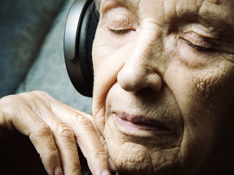
Why is it that we get chills when we listen to music? Why do our favorite old songs bring us right back to certain times in our life? In fact, this effect is seen even in those with Alzheimer’s disease. Researchers at the University of Utah Health examined the use of music in managing anxiety in individuals suffering from Anxiety. The study was discussed in a recent article in MedicalXpress:
"People with dementia are confronted by a world that is unfamiliar to them, which causes disorientation and anxiety" said Jeff Anderson, M.D., Ph.D., associate professor in Radiology at U of U Health and contributing author on the study. "We believe music will tap into the salience network of the brain that is still relatively functioning."
Previous work demonstrated the effect of a personalized music program on mood for dementia patients. This study set out to examine a mechanism that activates the attentional network in the salience region of the brain. The results offer a new way to approach anxiety, depression and agitation in patients with dementia. Activation of neighboring regions of the brain may also offer opportunities to delay the continued decline caused by the disease.
For three weeks, the researchers helped participants select meaningful songs and trained the patient and caregiver on how to use a portable media player loaded with the self-selected collection of music.
"When you put headphones on dementia patients and play familiar music, they come alive," said Jace King, a graduate student in the Brain Network Lab and first author on the paper. "Music is like an anchor, grounding the patient back in reality."
Using a functional MRI, the researchers scanned the patients to image the regions of the brain that lit up when they listened to 20-second clips of music versus silence. The researchers played eight clips of music from the patient's music collection, eight clips of the same music played in reverse and eight blocks of silence. The researchers compared the images from each scan.
The researchers found that music activates the brain, causing whole regions to communicate. By listening to the personal soundtrack, the visual network, the salience network, the executive network and the cerebellar and corticocerebellar network pairs all showed significantly higher functional connectivity.
"This is objective evidence from brain imaging that shows personally meaningful music is an alternative route for communicating with patients who have Alzheimer's disease," said Norman Foster, M.D., Director of the Center for Alzheimer's Care at U of U Health and senior author on the paper. "Language and visual memory pathways are damaged early as the disease progresses, but personalized music programs can activate the brain, especially for patients who are losing contact with their environment."
However, these results are by no means conclusive. The researchers note the small sample size (17 participants) for this study. In addition, the study only included a single imaging session for each patient. It is remains unclear whether the effects identified in this study persist beyond a brief period of stimulation or whether other areas of memory or mood are enhanced by changes in neural activation and connectivity for the long term.
"In our society, the diagnoses of dementia are snowballing and are taxing resources to the max," Anderson said. "No one says playing music will be a cure for Alzheimer's disease, but it might make the symptoms more manageable, decrease the cost of care and improve a patient's quality of life."
Read the original article Here
- Comments (0)
- Expressive Writing "Cools" The Worried Brain
- By Jason von Stietz, M.A.
- September 22, 2017
-

Getty Images It is often recommended that people write about their feelings. However, does writing calm a worried brain? Researchers at Michigan State University found that expressive writing frees cognitive resources and improves performance on a cognitive task. The study was discussed in a recent article in MedicalXpress:
The research, funded by the National Science Foundation and National Institutes of Health, provides the first neural evidence for the benefits of expressive writing, said lead author Hans Schroder, an MSU doctoral student in psychology and a clinical intern at Harvard Medical School's McLean Hospital.
"Worrying takes up cognitive resources; it's kind of like people who struggle with worry are constantly multitasking – they are doing one task and trying to monitor and suppress their worries at the same time," Schroder said. "Our findings show that if you get these worries out of your head through expressive writing, those cognitive resources are freed up to work toward the task you're completing and you become more efficient."
Schroder conducted the study at Michigan State with Jason Moser, associate professor of psychology and director of MSU's Clinical Psychophysiology Lab, and Tim Moran, a Spartan graduate who's now a research scientist at Emory University. The findings are published online in the journal Psychophysiology.
For the study, college students identified as chronically anxious through a validated screening measure completed a computer-based "flanker task" that measured their response accuracy and reaction times. Before the task, about half of the participants wrote about their deepest thoughts and feelings about the upcoming task for eight minutes; the other half, in the control condition, wrote about what they did the day before.
While the two groups performed at about the same level for speed and accuracy, the expressive-writing group performed the flanker task more efficiently, meaning they used fewer brain resources, measured with electroencephalography, or EEG, in the process.
Moser uses a car analogy to describe the effect. "Here, worried college students who wrote about their worries were able to offload these worries and run more like a brand new Prius," he said, "whereas the worried students who didn't offload their worries ran more like a '74 Impala – guzzling more brain gas to achieve the same outcomes on the task."
While much previous research has shown that expressive writing can help individuals process past traumas or stressful events, the current study suggests the same technique can help people – especially worriers – prepare for stressful tasks in the future.
"Expressive writing makes the mind work less hard on upcoming stressful tasks, which is what worriers often get "burned out" over, their worried minds working harder and hotter," Moser said. "This technique takes the edge off their brains so they can perform the task with a 'cooler head.'"
View the article Here
- Comments (0)
- Study Links Optimism, Anxiety, and Orbitifrontal Cortex Size
- By Jason von Stietz, M.A.
- October 3, 2015
-

Photo Credit: Getty Images Can optimism be traced to a structure in the brain? Researchers at the University of Illinois have linked optimism, anxiety, and the orbitofrontal cortex (OFC). Their study found that those with a larger OFC tend to report more optimism and less anxiety. The study was discussed in a recent article in MedicalXpress:
The new analysis, reported in the journal Social, Cognitive and Affective Neuroscience, offers the first evidence that optimism plays a mediating role in the relationship between the size of the OFC and anxiety.
Anxiety disorders afflict roughly 44 million people in the U.S. These disorders disrupt lives and cost an estimated $42 billion to $47 billion annually, scientists report.
The orbitofrontal cortex, a brain region located just behind the eyes, is known to play a role in anxiety. The OFC integrates intellectual and emotional information and is essential to behavioral regulation. Previous studies have found links between the size of a person's OFC and his or her susceptibility to anxiety. For example, in a well-known study of young adults whose brains were imaged before and after the colossal 2011 earthquake and tsunami in Japan, researchers discovered that the OFC actually shrank in some study subjects within four months of the disaster. Those with more OFC shrinkage were likely to also be diagnosed with post-traumatic stress disorder, the researchers found.
Other studies have shown that more optimistic people tend to be less anxious, and that optimistic thoughts increase OFC activity.
The team on the new study hypothesized that a larger OFC might act as a buffer against anxiety in part by boosting optimism.
Most studies of anxiety focus on those who have been diagnosed with anxiety disorders, said University of Illinois researcher Sanda Dolcos, who led the research with graduate student Yifan Hu and psychology professor Florin Dolcos. "We wanted to go in the opposite direction," she said. "If there can be shrinkage of the orbitofrontal cortex and that shrinkage is associated with anxiety disorders, what does it mean in healthy populations that have larger OFCs? Could that have a protective role?"
The researchers also wanted to know whether optimism was part of the mechanism linking larger OFC brain volumes to lesser anxiety.
The team collected MRIs of 61 healthy young adults and analyzed the structure of a number of regions in their brains, including the OFC. The researchers calculated the volume of gray matter in each brain region relative to the overall volume of the brain. The study subjects also completed tests that assessed their optimism and anxiety, depression symptoms, and positive (enthusiastic, interested) and negative (irritable, upset) affect.
A statistical analysis and modeling revealed that a thicker orbitofrontal cortex on the left side of the brain corresponded to higher optimism and less anxiety. The model also suggested that optimism played a mediating role in reducing anxiety in those with larger OFCs. Further analyses ruled out the role of other positive traits in reducing anxiety, and no other brain structures appeared to be involved in reducing anxiety by boosting optimism.
"You can say, 'OK, there is a relationship between the orbitofrontal cortex and anxiety. What do I do to reduce anxiety?'" Sanda Dolcos said. "And our model is saying, this is working partially through optimism. So optimism is one of the factors that can be targeted."
"Optimism has been investigated in social psychology for years. But somehow only recently did we start to look at functional and structural associations of this trait in the brain," Hu said. "We wanted to know: If we are consistently optimistic about life, would that leave a mark in the brain?"
Florin Dolcos said future studies should test whether optimism can be increased and anxiety reduced by training people in tasks that engage the orbitofrontal cortex, or by finding ways to boost optimism directly.
"If you can train people's responses, the theory is that over longer periods, their ability to control their responses on a moment-by-moment basis will eventually be embedded in their brain structure," he said.
Read the full article Here
- Comments (0)
- Brain Scans Predict Success of CBT for Social Anxiety Disorder
- By Jason von Stietz
- August 20, 2015
-
.jpg)
Photo Credit: MIT News Social anxiety disorder (SAD) affects 15 million people in the United States, approximately 6.8 percent of the U.S. population. Unfortunately, many treatments of SAD succeed as often as they fail. However, researchers at MIT found that brain scans predicted which patients would benefit from CBT with an accuracy of 80 percent. Medical Xpress discussed the findings in a recent article:
For patients with social anxiety disorder (SAD), current behavioral and pharmaceutical treatments work about half the time. After weeks of investment in therapy, about half of patients will likely still suffer with symptoms of anxiety, and have little choice but to try again with something else. This trial-and-error process—inevitable due to an absence of tools to guide treatment selection—is time-consuming and expensive, and some patients eventually just give up.
But new MIT research suggests that it may be possible to do better than a coin toss when choosing psychiatric therapies for patients. The study, which performed brain scans on 38 SAD patients, found that these scans contain clues that indicate, with about 80 percent accuracy, which SAD patients will do well in cognitive behavioral therapy (CBT), an intervention designed to help patients change thinking patterns. Use of the scans to predict treatment outcomes improved predictions fivefold over use of a clinician's assessment alone.
"Choice of therapy is like a wheel of chance," says first author Susan Whitfield-Gabrieli, a research scientist in the McGovern Institute for Brain Research at MIT. "We're hoping to use brain imaging to help provide more reliable predictors of treatment response."
The researchers used a form of brain imaging that scans patients in a state of rest. Resting-state images can be done quickly, in about 15 minutes, and reliably, since they don't require patients to follow instructions, so they have the potential to be used in a clinical setting as a tool that helps doctors select the best treatments for patients.
"Knowing who to give which therapy to upfront would save time, money, and health care resources," says Greg Siegle, an associate professor of psychiatry at the University of Pittsburgh School of Medicine who was not involved in this study. "This ability would be staggering to have at our disposal for the health care system."
The findings are reported in the current issue of the journal Molecular Psychiatry. The work was carried out in the lab of principal investigator John Gabrieli, the Grover Hermann Professor of Health Sciences and Cognitive Neuroscience at MIT and a member of the McGovern Institute.
Common disorder
Social anxiety disorder affects approximately 6.8 percent of Americans, about 15 million individuals, and is the country's third-most-common mental health disorder, according to the National Institutes of Mental Health. Its symptoms include extreme anxiety in social settings that can interfere with work and quality of life. Patients living with this disorder are also at higher risk of other psychiatric disorders, such as depression and substance abuse.
The study analyzed SAD patients from the Center for Anxiety and Related Disorders at Boston University and the Center for Anxiety and Traumatic Stress at Massachusetts General Hospital. The patients were scanned prior to participation in 12 weeks of group-based CBT. They also were evaluated using a behavioral assessment tool called the Liebowitz Social Anxiety Scale (LSAS) before and after CBT to determine who had improved.
In 2013, co-author Satrajit Ghosh, a principal research scientist at the McGovern Institute, studied task-based scans of this same group of patients. He and Whitfield-Gabrieli found that scans of patients' brains as they responded to angry or neutral faces and emotional or neutral scenes predicted CBT outcomes.
"But task-based scans have downsides," Whitfield-Gabrieli says: Behavioral differences among patients can affect performance. Also, task-based scans can only be used on patients who can follow a task, which excludes infants and some elderly or very ill patients.
Scanning the resting brain
So Whitfield-Gabrieli followed this earlier research with a study of the predictive power of resting-state imaging, which she and colleagues had also performed prior to CBT. During a resting-state scan, the patient just lies there. "That's the beauty of it," she says. "They're just letting their minds wander."
Resting-state imaging provides a look at the way a patient's brain is wired, both structurally and functionally. For instance, resting-state functional magnetic resonance imaging (fMRI) shows which parts of the brain synchronize with one another during rest, suggesting that they are functionally connected. In addition, analysis of diffusion-weighted magnetic resonance imaging (dMRI) shows the underlying anatomy of the white matter tracts that interconnect distant brain regions.
Based on findings from their earlier research, Whitfield-Gabrieli and colleagues first used resting-state fMRI to look at connections to the amygdala, the seat of fear in the brain. They found that patients with higher connectivity to the amygdala from certain other regions were more likely to have lower anxiety after CBT.
They then performed a second analysis of the resting-state fMRI data, this time looking across the entire brain for patterns of connectivity. This analysis revealed additional markers that were predictive of treatment. The researchers also analyzed dMRI data and found that more robust connectivity in the tract that connects visual cues with emotional responses is also predictive of improvement with CBT.
Higher LSAS scores, which indicate more severe SAD, correlate modestly with larger improvements after CBT. In this study, each brain scan analysis had predictive value beyond the LSAS, and the three analyses together produced a fivefold improvement in predictive power over the LSAS alone.
The next step for Whitfield-Gabrieli and colleagues is to validate their predictive model on hundreds or possibly thousands of patients. Such a large-scale study may be possible because resting-state scans are comparable even when performed in different labs or by different researchers. Such comparisons weren't feasible using task-based scans, which tend to vary from lab to lab.
"Right now there's a huge movement to create massive data sets, to share resting-state imaging data, and really change the way people do science," Whitfield-Gabrieli says.
Other next steps for Whitfield-Gabrieli's group are studies to predict the success of more than one form of therapy, and to look at other psychiatric conditions, such as depression and attention disorders. "We don't want to just know if they're going to respond to one treatment," she says. "We want to know which treatment is best for each patient."
Read the original article Here
- Comments (0)
- Brain Circuit Involved in Inheritance of Anxiety in Rhesus Monkeys
- By Jason von Stietz
- July 17, 2015
-

Photo Credit: Getty Images Anxious parents are likely to have anxious children. This is true of rhesus monkeys as well as humans. Researchers at the University of Wisconsin, Madison utilized positron emission topography (PET) scans to study anxiety in multiple generations of rhesus monkeys. Findings indicated that the inheritance of an over-active brain circuit might set the stage for the development of anxiety or depressive disorders. The study was discussed in a recent issue of NeuroScientistNews:
In rhesus monkey families -- just as in their human cousins -- anxious parents are more likely to have anxious offspring. And a new study in an extended family of monkeys provides important insights into how the risk of developing anxiety and depression is passed from parents to children.
The study from the Department of Psychiatry and the Health Emotions Research Institute at the University of Wisconsin-Madison shows how an over-active brain circuit involving three brain areas inherited from generation to generation may set the stage for developing anxiety and depressive disorders.
The study is published online in the early edition of the Proceedings of the National Academy of Sciences. It shows that elevated activity in this prefrontal- limbic -midbrain circuit is likely involved in mediating the in-born risk for extreme anxiety, anxious temperament that can be observed in early childhood.
"Over-activity of these three brain regions are inherited brain alterations that are directly linked to the later life risk to develop anxiety and depression,'' says senior author Dr. Ned Kalin, chair of psychiatry at the UW School of Medicine and Public Health. "This is a big step in understanding the neural underpinnings of inherited anxiety and begins to give us more selective targets for treatment."
Previous research by Kalin's group has shown that anxious temperament is inherited, and explained the brain circuits involved. About half of children who show extreme anxiety go on to develop stress-related psychiatric disorders later in life.
Monkeys, like humans, can be temperamentally anxious and pass their anxiety-related genes on to the next generation.
By studying nearly 600 young rhesus monkeys from a large multi-generational family, Drs. Andrew Fox, Kalin, and colleagues found that about 35 percent of variation in anxiety-like tendencies is explained by family history.
To understand which brain regions are responsible for passing anxiety from generation to generation, the authors measured anxiety-related behavior with high-resolution functional and structural brain imaging. They exposed the young monkeys to a mildly threatening situation that a child would also encounter, exposure to a stranger who does not make eye contact with the monkey. During this encounter, they used imaging methods commonly used in humans (positron emission tomography, PET) to identify brain regions in which increased metabolism predicted each individual's level of anxiety.
By closely examining how individual differences in brain function and anxiety-related behavior fall through the family tree, the authors identified brain systems responsible for the parent-to-child transmission of anxiety-related behavior. Using this "genetic correlation" approach, the authors found the neural circuit where metabolism and an early-life anxious temperament are likely to share the same genetic basis.
Interestingly, the brain circuit that was genetically correlated with individual differences in early-life anxiety involved three survival-related brain regions. These regions were located in the brain stem, the most primitive part of the brain; the amygdala, the limbic brain fear center; and the prefrontal cortex, which is responsible for higher-level reasoning and is fully developed only in humans and their primate cousins.
"Basically, we think that to a certain extent, anxiety can provide an evolutionary advantage because it helps an individual recognize and avoid danger, but when the circuits are over-active, it becomes a problem and can result in anxiety and depressive disorders," Kalin explains.
Surprisingly, these studies found that it was the function of these brain structures -- and not their size -- that was responsible for the genetic transfer of an anxious temperament. Although the search for the genetic underpinnings of anxiety have thus far been elusive, this research helps explain how genes might affect brain function and lead to extreme childhood anxiety, which greatly increases the risk to develop anxiety and depressive disorders.
"Now that we know where to look, we can develop a better understanding of the molecular alterations that give rise to anxiety-related brain function,'' Kalin says. "Our genes shape our brains to help make us who we are."
Read the original article Here
- Comments (0)
- tDCS and Attention Bias Modification Training Lead to Decreases in Anxiety
- By Jason von Stietz
- January 2, 2015
-
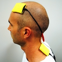
Photo Credit: University of Western Australia Researchers at Western University of Australia found that a combination of cognitive training and brain stimulation can lead to decreases in anxiety and depression. The study examined the use of cognitive bias modification and transcranial direct current stimulation in retraining maladaptive cognitive and attentional habits. The study was discussed in a recent article of MedicalXpress:
In collaboration with researchers at the University of Oxford, the study revealed that around 20 minutes of targeted electrical stimulation to a region of the frontal cortex could dramatically improve the effectiveness of a computer-based task designed to retrain unhelpful patterns of attention that are known to maintain high levels of anxiety.
Lead author, Dr Patrick Clarke, of UWA's School of Psychology and Centre for the Advancement of Research on Emotion, said the cognitive retraining procedure, known as attention bias modification, had shown considerable promise as a treatment foranxiety disorders, depression, addiction, and may even help with overeating.
"It works by having people practise a simple task where they have to repeatedly ignore certain unhelpful information, such as angry faces, or negative words, that would normally grab their attention," Dr Clarke said.
"The more the task can help people to direct their attention away from this type of unhelpful information, the more benefit they tend to get from it in terms of lower anxiety.
"Our Oxford colleagues were previously able to identify an area of the frontal cortexthat they believed could be responsible for the crucial change in attention that these tasks try to achieve. What's particularly exciting about our study is that we've been able to show that delivering electrical stimulation to this same area can enhance the effectiveness of the training."
The stimulation technique, known as transcranial direct current stimulation, or tDCS, can enhance activity in areas of the brain by applying a weak electrical current to the scalp.
"There has been some research looking into tDCS as a stand-alone treatment for conditions such as depression, but our findings suggest that it might be best used in conjunction with specific cognitive training tasks, such as the one we used," Dr Clarke said.
Read the original article Here
- Comments (1)
- Researchers Tie Social Behavior to Activity in Specific Brain Circuit
- By Jason von Stietz
- June 27, 2014
-

Photo Credit: Getty Images Can social behavior be linked back to specific wiring in the brain? Researchers at Stanford University examined the relationships between a specific circuit in the brain and the tendency of mammals to interact socially. When researchers stimulated this circuit in mice they instantly engaged in social behavior with unfamiliar mice. In contrast, when researchers inhibited this circuit the mice refrained from social activity and ignored the other mice. Medical Xpress discussed the study in a recent article:
The new findings, to be published June 19 inCell, may throw light on psychiatric disorders marked by impaired social interaction such as autism, social anxiety, schizophrenia and depression, said the study's senior author, Karl Deisseroth, MD, PhD, a professor of bioengineering and of psychiatry and behavioral sciences. The findings are also significant in that they highlight not merely the role of one or another brain chemical, as pharmacological studies tend to do, but rather the specific components of brain circuits involved in a complex behavior. A combination of cutting-edge techniques developed in Deisseroth's laboratory permitted unprecedented analysis of how brain activity controls behavior.
Deisseroth, the D.H. Chen Professor and a member of the interdisciplinary Stanford Bio-X institute, is a practicing psychiatrist who sees patients with severe social deficits. "People with autism, for example, often have an outright aversion to social interaction," he said. They can find socializing—even mere eye contact—painful.
Deisseroth pioneered a brain-exploration technique, optogenetics, that involves selectively introducing light-receptor molecules to the surfaces of particular nerve cellsin a living animal's brain and then carefully positioning, near the circuit in question, the tip of a lengthy, ultra-thin optical fiber (connected to a laser diode at the other end) so that the photosensitive cells and the circuits they compose can be remotely stimulated or inhibited at the turn of a light switch while the animal remains free to move around in its cage.
Using optogenetics and other methods he and his associates have invented, Deisseroth and his associates were able to both manipulate and monitor activity in specific nerve-cell clusters, and the fiber tracts connecting them, in mice's brains in real time while the animals were exposed to either murine newcomers or inanimate objects in various laboratory environments. The mice's behavioral responses were captured by video and compared with simultaneously recorded brain-circuit activity.
In some cases, the researchers observed activity in various brain centers and nerve-fiber tracts connecting them as the mice variously examined or ignored one another. Other experiments involved stimulating or inhibiting impulses within those circuits to see how these manipulations affected the mice's social behavior.
To avoid confusing simple social interactions with mating- and aggression-related behaviors, the researchers restricted their experiments to female mouse pairs.
The scientists first examined the relationship between the mice's social interactions and a region in the brain stem called the ventral tegmental area. The VTA is a key node in the brain's reward circuitry, which produces sensations of pleasure in response to success in such survival-improving activities as eating, mating or finding a warm shelter in a cold environment.
The VTA transmits signals to other centers throughout the brain via tracts of fibers that secrete chemicals, including one called dopamine, at contact points abutting nerve cells within these faraway centers. When dopamine lands on receptors on those nerve cells, it can set off signaling activity within them.
Abnormal activity in the VTA has been linked to drug abuse and depression, for example. But much less is known about this brain center's role in social behavior, and it had not previously been possible to observe or control activity along its connections during social behavior.
Deisseroth and his colleagues used mice whose dopamine-secreting, or dopaminergic, VTA nerve cells had been bioengineered to express optogenetic control proteins that could set off or inhibit signaling in the cells in response to light. They observed that enhancing activity in these cells increased a mouse's penchant for social interaction. When a newcomer was introduced into its cage, it came, it saw, it sniffed. Inhibiting the dopaminergic VTA cells had the opposite effect: The host lost much of its interest in the guest.
Read the Full Article Here
- Comments (1)
- Brain Games: Move Objects with Your Mind to Find Inner Calm?
- By Jason von Stietz
- March 6, 2014
-
Couch potatoes everywhere, rejoice.
New commercial devices, using technology borrowed from the field of neuroscience, are making it possible to control objects with brain power alone. The idea is to help train users to become more focused — and relaxed. Read Full Article Here
- Comments (0)
 Subscribe to our Feed via RSS