Blog
- New Imaging Tool Measures Synaptic Density
- By Jason von Stietz, M.A.
- July 29, 2016
-
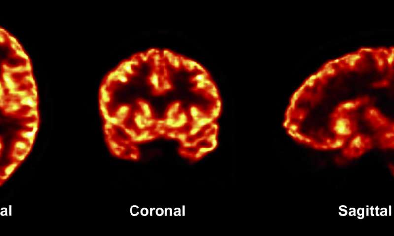
Photo Credit: Finemma et al. (2016). Science Translational Medicine Previously researcher have only been able to study the synaptic changes caused by brain disorders through autopsy. However, recently researcher from Yale have developed a new approach to brain scanning that allows for the measurement synaptic density. The study was discussed in a recent article in Medical Xpress:
The study was published July 20 in Science Translational Medicine.
Certain changes in synapses—the junctions between nerve cells in the brain—have been linked with brain disorders. But researchers have only been able to evaluate synaptic changes during autopsies. For their study, the research team set out to develop a method for measuring the number of synapses, or synaptic density, in the living brain.
To quantify synapses throughout the brain, professor of radiology and biomedical imaging Richard Carson and his coauthors combined PET scanning technology with biochemistry. They developed a radioactive tracerthat, when injected into the body, binds with a key protein that is present in all synapses across the brain. They observed the tracer through PET imaging and then applied mathematical tools to quantify synaptic density.
The researchers used the imaging technique in both baboons and humans. They confirmed that the new method did serve as a marker for synaptic density. It also revealed synaptic loss in three patients with epilepsy compared to healthy individuals.
"This is the first time we have synaptic density measurement in live human beings," said Carson, who is senior author on the study. "Up to now any measurement of synaptic density was postmortem."
The finding has several potential applications. With this noninvasive method, researchers may be able to follow the progression of many brain disorders, including epilepsy and Alzheimer's disease, by measuring changes in synaptic density over time. Another application may be in assessing how pharmaceuticals slow the loss of neurons. "This opens the door to follow the natural evolution of synaptic density with normal aging and follow how drugs can alter synapses or synapse formation."
Carson and his colleagues plan future studies involving PET imaging of synapses to research epilepsy and other brain disorders, including Alzheimer's disease, schizophrenia, depression, and Parkinson's disease. "There are many diseases where neuro-degeneration comes into play," he noted.
Read the original article Here
- Comments (0)
- Impact Of Baby's Cries on Cognition
- By Jason von Stietz, M.A.
- July 20, 2016
-

Getty Images People often refer to “parental instincts” as an innate drive to care for one’s offspring. However, we know little of the role cognition play in this instinct. Researchers at the University of Toronto examined the impact of the sound of a baby’s cries on performance during a cognitive task. The study was discussed in a recent article in Neuroscience Stuff:
“Parental instinct appears to be hardwired, yet no one talks about how this instinct might include cognition,” says David Haley, co-author and Associate Professor of psychology at U of T Scarborough.
“If we simply had an automatic response every time a baby started crying, how would we think about competing concerns in the environment or how best to respond to a baby’s distress?”
The study looked at the effect infant vocalizations—in this case audio clips of a baby laughing or crying—had on adults completing a cognitive conflict task. The researchers used the Stroop task, in which participants were asked to rapidly identify the color of a printed word while ignoring the meaning of the word itself. Brain activity was measured using electroencephalography (EEG) during each trial of the cognitive task, which took place immediately after a two-second audio clip of an infant vocalization.
The brain data revealed that the infant cries reduced attention to the task and triggered greater cognitive conflict processing than the infant laughs. Cognitive conflict processing is important because it controls attention—one of the most basic executive functions needed to complete a task or make a decision, notes Haley, who runs U of T’s Parent-Infant Research Lab.
“Parents are constantly making a variety of everyday decisions and have competing demands on their attention,” says Joanna Dudek, a graduate student in Haley’s Parent-Infant Research Lab and the lead author of the study.
“They may be in the middle of doing chores when the doorbell rings and their child starts to cry. How do they stay calm, cool and collected, and how do they know when to drop what they’re doing and pick up the child?”
A baby’s cry has been shown to cause aversion in adults, but it could also create an adaptive response by “switching on” the cognitive control parents use in effectively responding to their child’s emotional needs while also addressing other demands in everyday life, adds Haley.
“If an infant’s cry activates cognitive conflict in the brain, it could also be teaching parents how to focus their attention more selectively,” he says.
“It’s this cognitive flexibility that allows parents to rapidly switch between responding to their baby’s distress and other competing demands in their lives—which, paradoxically, may mean ignoring the infant momentarily.”
The findings add to a growing body of research suggesting that infants occupy a privileged status in our neurobiological programming, one deeply rooted in our evolutionary past. But, as Haley notes, it also reveals an important adaptive cognitive function in the human brain.
Read the original article here
- Comments (0)
- Hypoconnectivity Found In Brains Of Those With Intermittent Explosive Disorder
- By Jason von Stietz, M.A.
- July 15, 2016
-

Photo Credit: Lee et al., Neuropsychopharmacology Why do those with anger issues tend to misunderstand social situations? Researchers at the University of Chicago Medical School compared the brains of those suffering from intermittent explosive disorder (IED) to the brains of healthy controls using diffusion tensor imaging. Findings indicated that brains of people with IED showed less white matter connecting the frontal cortex to the parietal lobes. The study was discussed in a recent article in Medical Xpress:
In a new study published in the journal Neuropsychopharmacology, neuroscientists from the University of Chicago show that white matter in a region of the brain called the superior longitudinal fasciculus (SLF) has less integrity and density in people with IED than in healthy individuals and those with other psychiatric disorders. The SLF connects the brain's frontal lobe—responsible for decision-making, emotion and understanding consequences of actions—with the parietal lobe, which processes language and sensory input.
"It's like an information superhighway connecting the frontal cortex to the parietal lobes," said Royce Lee, MD, associate professor of psychiatry and behavioral neuroscience at the University of Chicago and lead author of the study. "We think that points to social cognition as an important area to think about for people with anger problems."
Lee and his colleagues, including senior author Emil Coccaro, MD, Ellen C. Manning Professor and Chair of Psychiatry and Behavioral Neuroscience at UChicago, used diffusion tensor imaging, a form ofmagnetic resonance imaging (MRI) that measures the volume and density of white matter connective tissue in the brain. Connectivity is a critical issue because the brains of people with psychiatric disorders usually show very few physical differences from healthy individuals.
"It's not so much how the brain is structured, but the way these regions are connected to each other," Lee said. "That might be where we're going to see a lot of the problems in psychiatric disorders, so white matter is a natural place to start since that's the brain's natural wiring from one region to another."
People with anger issues tend to misunderstand the intentions of other people in social situations. They think others are being hostile when they are not and make the wrong conclusions about their intentions. They also don't take in all the data from a social interaction, such as body language or certain words, and notice only those things that reinforce their belief that the other person is challenging them.
Decreased connectivity between regions of the brain that process a social situation could lead to the impaired judgment that escalates to an explosive outburst of anger. The discovery of connectivity deficits in a specific region of the brain like the SLF provides an important starting point for more research on people with IED, as well as those with borderline personality disorder, who share similar social and emotional problems and appear to have the same abnormality in the SLF.
"This is another example of tangible deficits in the brains of those with IED that indicate that impulsive aggressive behavior is not simply 'bad behavior' but behavior with a real biological basis that can be studied and treated," Coccaro said.
Read the original article Here
- Comments (0)
 Subscribe to our Feed via RSS