Blog
- Cognitive Training Reduces Depression
- By Jason von Stietz, M.A.
- June 30, 2018
-
.jpg)
iStock About half of individuals who suffer a traumatic brain injury (TBI) develop depression within a year. However, would a treatment designed to address cognitive symptoms also reduce depression? Researchers from the Center for Brain Health at the University of Texas at Dallas examined the use of cognitive training programs to address depression in those who suffered a TBI. Findings indicated that cognitive training, even when not directed toward psychiatric symptoms, related to not only cognitive improvements but also reductions in depression. The study was discussed in a recent article in Medical Xpress:
The recent study, published in Human Brain Mapping, revealed significant reductions in the severity of depressive symptoms, increased ability to regulate emotions, increases in cortical thickness and recovery from abnormal neural network connectivity after cognitive training.
"To our knowledge, this is the first study to report brain change associated with reduced depression symptoms after cognitive training," said Dr. Kihwan Han, a research scientist at the Center for BrainHealth who works in the lab of Dr. Daniel Krawczyk. Han is the lead author of the study.
"Overall, these findings suggest that cognitive training can reduce depressive symptoms in patients with traumatic brain injury even when the training does not directly target psychiatric symptoms," he said.
A past study involving the same protocol showed cognitive gains as well as similar changes in cortical thickness and neural network connectivity.
This study included 79 individuals with chronic TBI who all were at least six months post-injury. These individuals were randomly assigned into one of two groups: strategy-based training, which used the Strategic Memory Advanced Reasoning Training (SMART) program developed at the center; and information-based training, which used the Brain Health Workshop program. Researchers used the Beck Depressive Inventory to classify 53 of the participants as depressed.
The participants' depressive-symptom severity, psychological functioning scores and data from magnetic resonance imaging brain scans were collected before training, after training and three months post-training. Scans were used to study changes in brain structure and neural network connectivity.
Both training programs consisted of 12 one-and-a half-hour sessions over eight weeks that included quizzes, homework assignments, and projects conducted in small group settings that involved social interactions.
All participants in the depressed group showed significantly reduced depressive symptoms that were associated with improvements in cognitive and daily life functioning. According to Han, the social engagements, cognitive stimulation from new learning opportunities and hope of improvement afforded by both programs may help explain the reductions in depressive symptoms.
Based on the observed brain change patterns, Han suggested that improved emotion regulation also may be related to the reduced depressive symptoms. Over time, the reductions in depression symptom severity correlated with increased cortical thickness within the prefrontal cortex—an area of the brain responsible for executive functions needed for emotional control—and reductions in abnormally elevated neural connectivity within this region.
"Identifying what changes are happening in the brain when interventions successfully reduce depressive symptoms could allow us to create more effective, pharmaceutical-free approaches to help alleviate depression in people who experience chronic traumatic brain injury symptoms," said study author Dr. Sandra Bond Chapman, founder and chief director of the Center for BrainHealth, and Dee Wyly Distinguished University Professor.
Read the original article Here
- Comments (0)
- Gist Reasoning and Traumatic Brain Injury
- By Jason von Stietz, M.A.
- February 24, 2017
-

BigStock The ability to understand the “gist” of things is an important and likely underappreciated skill. Gist reasoning, or the ability to look at complex, concrete information and make deeper level abstract interpretations is an essential part of each person’s daily activities. Researchers at Texas Woman’s University and University of Texas Dallas studied the relationship between gist reasoning and cognitive deficits in individual’s with traumatic brain injury. The study was discussed in a recent article in Medical Xpress:
The study, published in Journal of Applied Biobehavioral Research, suggests the gist reasoning test may be sensitive enough to help doctors and clinicians identify previously undiagnosed cognitive changes that could explain the daily life difficulties experienced by TBI patients and subsequently guide appropriate therapies.
The gist reasoning measure, called the Test of Strategic Learning, accurately identified 84.7 percent of chronic TBI cases, a much higher rate than more traditional tests that accurately identified TBI between 42.3 percent and 67.5 percent of the time.
"Being able to 'get the gist' is essential for many day-to-day activities such as engaging in conversation, understanding meanings that are implied but not explicitly stated, creating shopping lists and resolving conflicts with others," said study lead author Dr. Asha Vas of Texas Woman's University who was a postdoctoral fellow at the Center for BrainHealth at the time of the study. "The gist test requires multiple cognitive functions to work together."
The study featured 70 participants ages 18 to 55, including 30 who had experienced a moderate to severe chronic traumatic brain injury at least one year ago. All the participants had similar socioeconomic status, educational backgrounds and IQ.
Researchers were blinded to the participant's TBI status while administering four different tests that measure abstract thinking—the ability to understand the big picture, not just recount the details of a story or other complex information. Researchers used the results to predict which participants were in the TBI group and which were healthy controls.
During the cognitive tests, the majority of the TBI group easily recognized abstract or concrete information when given prompts in a yes-no format. But the TBI group performed much worse than controls on tests, including gist reasoning, that required deeper level processing of information with fewer or no prompts.
The gist reasoning test consists of three texts that vary in length (from 291 to 575 words) and complexity. The test requires the participant to provide a synopsis of each of the three texts.
Vas provided an example of what "getting the gist" means using Shakespeare's play Romeo and Juliet.
"There are no right or wrong answers. The test relies on your ability to derive meaning from important story details and arrive at a high-level summary: Two young lovers from rival families scheme to build a life together and it ends tragically. You integrate existing knowledge, such as the concept of love and sacrifice, to create a meaning from your perspective. Perhaps, in this case, 'true love does not conquer all,'" she said.
Past studies have shown that higher scores on the gist reasoning test in individuals in chronic phases of TBI correlate to better ability to perform daily life functions.
"Perhaps, in the future, the gist reasoning test could be used as a tool to identify other cognitive impairments," said Dr. Jeffrey Spence, study co-author and director of biostatistics at the Center for BrainHealth. "It may also have the potential to be used as a marker of cognitive changes in aging."
Read the original article Here
- Comments (0)
- Long-Term Effects of TBI Studied
- By Jason von Stietz, M.A.
- February 19, 2017
-

Getty Images The long-term effects of traumatic brain injury (TBI) are just now beginning to be understood. Researchers from Cincinnati Children’s Hospital Medical Center examined the impact of TBI on pediatric patients about seven years after the injury. The findings indicated that children who have suffered a mild to moderate TBI are twice as likely to have issues with attention than their healthy counterparts. The study was discussed in a recent issue in Medical Xpress:
In a study to be presented Friday Feb. 10 at the annual meeting of the Association of Academic Physiatrists in Las Vegas, researchers from Cincinnati Children's will present research on long-term effects of TBI—an average of seven years after injury. Patients with mild to moderate brain injuries are two times more likely to have developed attention problems, and those with severe injuries are five times more likely to develop secondary ADHD. These researchers are also finding that the family environment influences the development of these attention problems.
- Parenting and the home environment exert a powerful influence on recovery. Children with severe TBI in optimal environments may show few effects of their injuries while children with milder injuries from disadvantaged or chaotic homes often demonstrate persistent problems.
- Early family response may be particularly important for long-term outcomes suggesting that working to promote effective parenting may be an important early intervention.
- Certain skills that can affect social functioning, such as speed of information processing, inhibition, and reasoning, show greater long-term effects.
- Many children do very well long-term after brain injury and most do not have across the board deficits.
More than 630,000 children and teenagers in the United States are treated in emergency rooms for TBI each year. But predictors of recovery following TBI, particularly the roles of genes and environment, are unclear. These environmental factors include family functioning, parenting practices, home environment, and socioeconomic status. Researchers at Cincinnati Children's are working to identify genes important to recovery after TBI and understand how these genes may interact with environmental factors to influence recovery.
- They will be collecting salivary DNA samples from more than 330 children participating in the Approaches and Decisions in Acute Pediatric TBI Trial.
- The primary outcome will be global functioning at 3, 6, and 12 months post injury, and secondary outcomes will include a comprehensive assessment of cognitive and behavioral functioning at 12 months post injury.
- This project will provide information to inform individualized prognosis and treatment plans.
Using neuroimaging and other technologies, scientists are also learning more about brain structure and connectivity related to persistent symptoms after TBI. In a not-yet-published Cincinnati Children's study, for example, researchers investigated the structural connectivity of brain networks following aerobic training. The recovery of structural connectivity they discovered suggests that aerobic training may lead to improvement in symptoms.
Over the past two decades, investigators at Cincinnati Children's have conducted a series of studies to develop and test interventions to improve cognitive and behavioral outcomes following pediatric brain injury. They developed an innovative web-based program that provides family-centered training in problem-solving, communication, and self-regulation.
- Across a series of randomized trials, online family problem-solving treatment has been shown to reduce behavior problems and executive dysfunction (management of cognitive processes) in older children with TBI, and over the longer-term improved everyday functioning in 12-17 year olds.
- Web-based parenting skills programs targeting younger children have resulted in improved parent-child interactions and reduced behavior problems. In a computerized pilot trial of attention and memory, children had improvements in sustained attention and parent-reported executive function behaviors. These intervention studies suggest several avenues for working to improve short- and long-term recovery following TBI.
Read the original article Here
- Comments (0)
- Rest is Critical Following a Concussion
- By Jason von Stietz, M.A.
- February 27, 2016
-
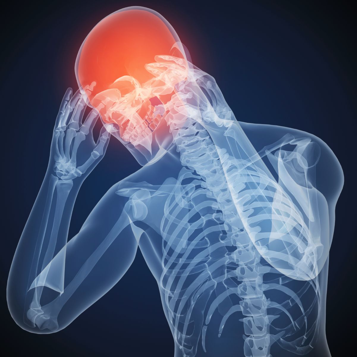
Photo Credit: Unknown Researchers at Georgetown University Medical Center (GUMC) found that resting after a concussion is of critical importance to restoring brain health. Using an animal model, researchers investigated the impact of weekly versus daily mild traumatic brain injuries in mice. Findings indicated that neuronal connections lost after an injury were recovered within three days. However, when insults to the brain were daily the neuronal connections were not restored and the impact of the injuries were detected in 1-year follow-up. The study was discussed in a recent article in a recent GUMC press release:
Doctors who order several days of rest after a person suffers a concussion are giving sound advice, say researchers, and new data from animal models explains why.
Georgetown University Medical Center neuroscientists say rest — for more than a day — is critical for allowing the brain to reset neural networks and repair any short-term injury. The new study in mice also shows that repeated mild concussions with only a day to recover between injuries leads to mounting damage and brain inflammation that remains evident a year after injury.
“It is good news that the brain can recover from a hit if given enough time to rest and recover. But on the flip side, we find that the brain does not undertake this rebalancing when impacts come too close together,” says the study’s lead researcher, Mark P. Burns, PhD, assistant professor of neuroscience at GUMC and director of the Laboratory for Brain Injury and Dementia.
This first-of-its-kind study, published in the March 2016 issue of American Journal of Pathology, modeled repeated mild head trauma as a means to investigate brain damage that occurs after a sports, military or domestic abuse injury.
Investigators developed a mouse model of repetitive, extremely mild concussive impacts conducted while the mouse is anesthetized. They compared the brain’s response to a single concussion with an injury received daily for 30 days and one received weekly over 30 weeks.
Mice with a single insult temporarily lose 10-15 percent of the neuronal connections in their brains, but no inflammation or cell death resulted, Burns says. With three days rest, all neuronal connections were restored. This neuronal response is not seen in mice with daily concussions, but the pattern is restored when a week of rest is given between each insult, Burns says.
When a mild concussion occurred each day for a month, inflammation and damage to the brain’s white matter resulted. “This damage became progressively worse for two months and remained apparent one year after the last impact,” Burns says.
“The findings mirror what has been observed about such damage in humans years after a brain injury, especially among athletes,” Burns says. “Studies have shown that almost all people with single concussions spontaneously recover, but athletes who play contact sports are much more susceptible to lasting brain damage. These findings help fill in the picture of how and when concussions and mild head trauma can lead to sustained brain damage.”
Georgetown co-authors are first author Charisse N. Winston, PhD, Maia Parsadanian, David N. Zapple, Sonia Villapol, PhD, and undergraduate students David Barton, Tiffany E. Wilkins, Aidan Neustadtl, Deepa Chellappa, and Andrew D. Alikhani. Contributors also include Emmanuel Planel, PhD, and Anastasia Noel, PhD, from the Centre Hospitalier de l'Université Laval, Neurosciences, Québec, Canada.
The study was supported by Georgetown University’s Neural Injury and Plasticity Training Program, the National Institute for Neurological Disorders and Stroke (R01 NS067417), a supplement to Promote Diversity in Health-Related Research, a donation from KPB Corporation, the Canadian Institute of Health Research, Fonds de Recherche en Santé du Québec and the Natural Sciences and Engineering Research Council of Canada.
Read the oringinal article Here
- Comments (0)
- Surgical Treatment of TBI in Older Adults
- By Jason von Stietz, M.A.
- November 30, 2015
-

Getty Images Researches at the Helsinki University Hospital Department of Neurosurgery investigated these use of surgery to treat acute subdural hemotomas in patients over the age of 75. Previously, older adult patients were not treated surgically, as the rates of patents who survived and recovered successfully were low. However, new findings show that older adult patients who lived independently before the accident, were not taking anticoagulants, and who arrived at the hospital conscious received were treated successfully through surgery. The study was discussed in a recent article in NeuroScientistNews:
According to a study completed at the Helsinki University Hospital Department of Neurosurgery, even patients over the age of 75 may recover from severe traumatic brain injury. This is the first study to describe the results of surgically treated elderly patients with acute subdural hematomas.
It is generally accepted that elderly patients who suffer from an acute subdural hematoma should not be treated surgically, as few survive and even fewer recover to an independent life. However, the world's population is rapidly aging leading to an increased rate of fall accidents. In the worst case, falling may result in brain hemorrhage.
Age is one of the most significant outcome predictors in patients with traumatic brain injury. If the patient is young, an acute subdural hematoma is normally treated through a neurosurgical operation. However, even among young patients, mortality and significant morbidity are highly common, despite surgical treatment. In older patients, the success rate of the surgery are made worse by the fact that many patients are typically using oral anticoagulant medications to treat other cardiovascular diseases.
The Neurosurgical Department in Helsinki University Hospital has been an exception in its policy to also treat elderly patients with acute subdural hematomas surgically. Researchers from the University of Helsinki and Helsinki University Hospital have now determined how the patients' functional status before the injury and the use of oral anticoagulant medications influence the prognosis of patients 75 years or older operated on for an acute subdural hematoma.
The study showed that no patients who had been brought to hospital unconscious, who had not been independent before the trauma, or who had used anticoagulants were alive at one year after the surgery.
"What was surprising, however, was that patients who were conscious at presentation, who were not using anticoagulants or were independent before the operation, recovered quite well. The expected lifespan of these patients was comparable to their age-matched peers," says MD, PhD Rahul Raj, one of the main authors.
"One should be careful to make to strong conclusions from such a small number of patients," Raj points out, "but it seems that in approximately half of all cases, even elderly patients may benefit from surgery and recover to an independent life. It is important to note that included patients had an isolated acute subdural hematoma with no injuries to the brain tissue itself. This means that the results cannot be applied to patients with contusions or other intracranial injuries, whose treatment and prognosis are different."
The decision to operate should not be based on age alone
According to Raj, the study throws new light on the old assumption that surgical treatment of the elderly is not a sensible course of action: "The decision to treat through surgery should not be based on age alone, even though this is common."
Surgery of an acute subdural hematoma followed by intensive care and rehabilitation involve major costs and can cause significant suffering to patients and relatives. Thus, it is important to perform surgery on only the patients who are likely to benefit from it.
"But how do you define a bad prognosis? If only one in ten patients recovers sufficiently to live at home, is the treatment worthwhile? If half of the treated patients die within the year, is the treatment worthwhile? This is not a medical decision," the researchers emphasize. They believe that in the future, surgical treatment will be increasingly restricted to patients with the highest likelihood of recovering.
Read the original article Here
- Comments (2)
- Post-Concussion Syndrome Symptoms Linked to Inflammation
- By Jason von Stietz
- May 15, 2015
-

Photo Credit: Getty Images Why do people who have had a very mild head injury, or no head injury at all, but an injury to another part of the body, sometimes display symptoms of post concussion-like syndromes? Researchers at McMAster University have linked these symptoms to inflammation rather than concussions. The finding was discussed in a recent article in MedicalXpress:
A team of researchers based at McMaster University has developed a new understanding of post-concussion syndrome, answering questions that have been plaguing researchers in the field.
Their study, published in the medical journalBrain, Behavior and Immunity, provides an explanation for why many people with even very trivial head injuries, or even injuries to other parts of their bodies, experience incapacitating post-concussion like syndromes.
These symptoms include headaches, dizziness, cognitive impairment and otherneuropsychiatric symptoms such as irritability, anxiety and insomnia.
"It's inflammation that they have in common," said Michel Rathbone, a professor of medicine for McMaster's Michael G. DeGroote School of Medicine and a lead author of the paper. "Rather than a concussion, we'd like to propose a unifying umbrella term of post-inflammatory brain syndromes or PIBS."
He added that the research will encourage scientists to open up new lines of research into understanding the cause of post-concussion symptoms in the absence of obviously visible brain injury on conventional imaging and into the treatment of these symptoms by targeting inflammatory mediators. For example, people who have a very subtle genetic change in a certain inflammatory protein have poorer recovery after brain injury.
It also explains why many social factors appear to play a role in development ofsymptoms: "We know that the immune system can be modulated, or sensitized by the current and even the previous environment an individual was in. These social factors, such as preexisting stressors, depression or anxiety, may actually be, in a way, biological factors."
Rathbone added that this will provide hope for individuals with cognitive dysfunction after major infections, surgeries and traumas, as it suggests that current and future treatments for concussion may hold a benefit for these individuals.
"This research opens many doors for so many different patients. We are excited to be starting a totally new approach to the field, and we look forward to making a difference for the patients of the future."
Read the original article Here
- Comments (0)
- Is Neurofeedback Effective? The Washington Post Reviews Clinical Work, Research, and Personal Experiences
- By Jason von Stietz
- January 30, 2015
-
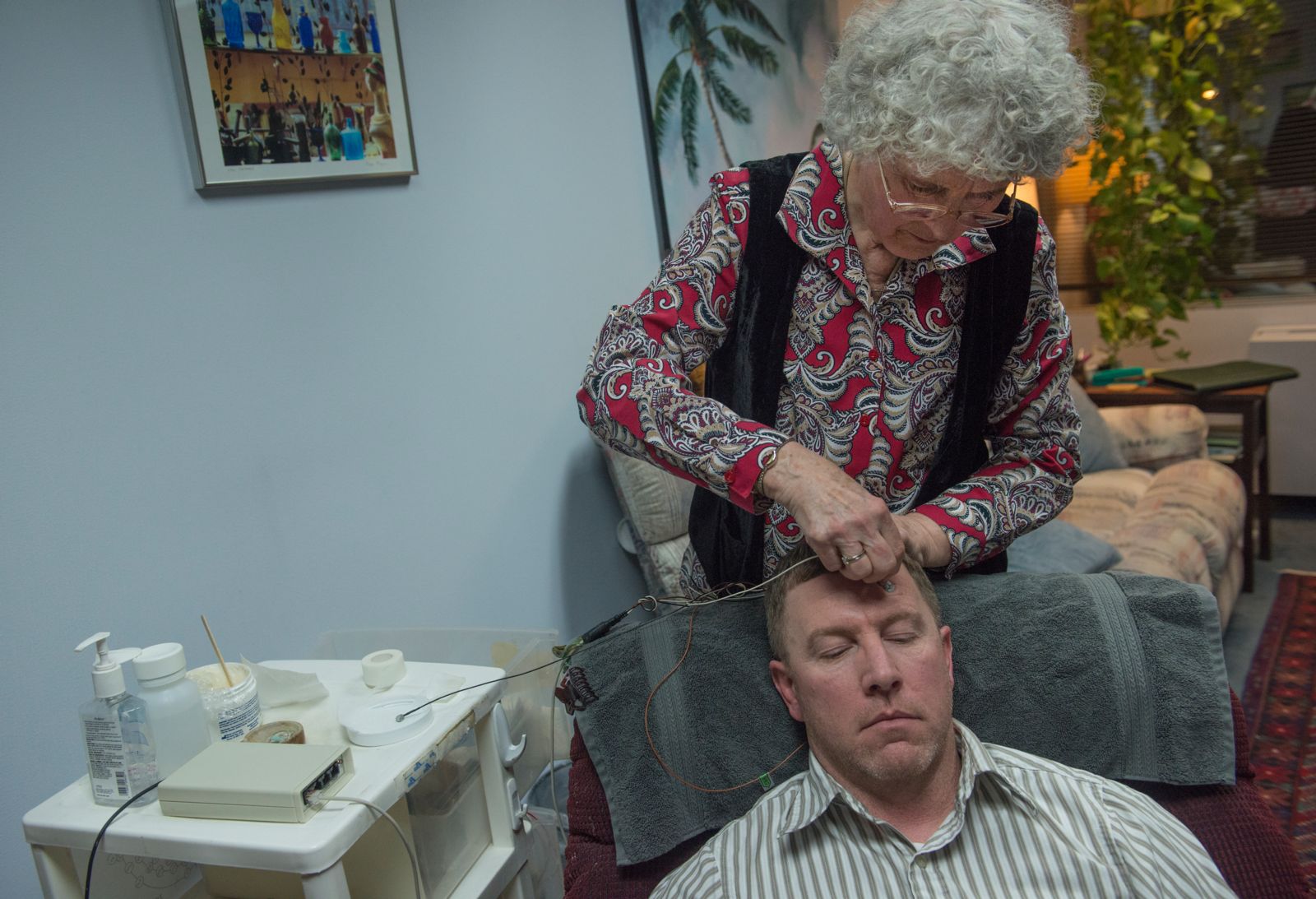
Photo Credit: Washington Post The media shined a light on the neurofeedback field in a recent article in the Washington Post. Many clinicians have developed expertise treating a variety of psychiatric disorders. Some researchers are unsure there is enough evidence to support the claims of neurofeedback practitioners. However, leading neurofeedback researchers point to the field’s recent studies as evidence. Arlene Karadis discussed the clinical work and research of neurofeedback experts and the personal experiences of their patients in a recent article:
In September 2013, Chris Gardner went from kicking and spinning as a black belt in taekwondo to being locked in a world where he could not follow conversations — or even walk his dog. The 58-year-old Vienna, Va., resident had just had brain surgery to remove a large tumor, and the operation affected his mobility and cognition.
After nine months of physical and occupational therapy, he’d made little progress. So he tried neurofeedback, hoping this controversial treatment would improve his balance and mental processes.
Neurofeedback — a type of biofeedback — uses movies, video games, computers and other tools to help individuals regulate their brain waves. A patient might watch a movie, for example, while hooked to sensors that send data to a computer. A therapist, following the brain activity on a monitor, programs the computer to stop the movie if an abnormal number of fast or slow brain waves is detected or if the brain waves are erratic, moving rapidly from fast to slow waves.
The stop-and-start feedback, repeated over and over in numerous sessions, seems to yield more-normal brain waves. Researchers who endorse the technique say they don’t know exactly how it works but they say the changes in brain waves result in improved ability to focus and relax.
Neurofeedback, which is also used for post-traumatic stress disorder and attention-deficit hyperactivity disorder, has been around since the 1960s. Some research has found it promising. Other studies have been inconclusive, and some have shown no positive outcomes.
The most solid data concern ADHD, especially a recent trial involving 104 children published in March in the Journal of Pediatrics. Those who received neurofeedback had improvements in attention and impulse control, while those who did not receive the therapy did not. These improvements persisted after six months. The authors concluded that neurofeedback may be a “promising attention training treatment for children with ADHD.”
Gardner had read that the technique could aid in recovery from brain injuries.
“I was skeptical. But I was desperate. I felt like I was wrapped in miles of cotton and could not reach through it to touch or feel anything,” said Gardner, an electronic technology consultant. His doctor was projecting a two-to-three-year recovery period, based on Gardner’s slow progress nine months after surgery.
By his ninth neurofeedback session, he was driving, taking power walks and working from home.
Neurofeedback treatments vary. In Gardner’s case, he sat in a chair while tiny, pulsed signals were sent to his brain. Research suggests that these signals enable the brain to revive its communication channels, which can become impaired after a brain injury.
“I couldn’t feel anything” while the treatments were underway, Gardner said. “I just sat there with my eyes closed. My therapist explained that the pulses basically reboot the brain.”
Better focus and relaxation can seemingly help improve or eliminate such conditions as migraines (imbalanced brain waves are associated with certain symptoms like pain) and anxiety.
He has just completed the last of 10 treatments. “I am not 100 percent. I probably won’t stand on my head or get on a roller coaster. But I can do almost everything I couldn’t do before,” said Gardner, who’s back to his martial arts.
“Do most people become totally normal? No. But they improve,” said Michael Sitar, a Bethesda psychologist certified in neurofeedback. He uses it to treat depression, ADHD, chronic pain and some other conditions.
“I find [that] people with focus problems can switch tasks easier. Kids who repeat themselves and who are emotionally labile become calmer and don’t repeat as much,” Sitar said. “With some complicated cases, like bipolar disorder, people may get by on less medication. Though less common, there are documented cases of nonverbal people who become verbal.”
Like riding a bike
Deborah Stokes, an Alexandria psychologist, compares neurofeedback to riding a bike: It’s non-conscious learning, based on the feedback, that, with repetition, can be long-lasting, she said.
“We don’t know exactly how neurofeedback works,” she said. “It’s a process where if clients get out of their own way, they relax. Over time, they get the desired brain pattern, feel calm and function better. This encourages them to stay with it.” Her team sees 30 patients a week.
Thomas Nicklin, whose family was living in Alexandria, saw Stokes for debilitating migraines. A year and a half after beginning a drug regimen prescribed by a neurologist, he was not getting better.
Nicklin, a teenager who was in boarding school, did 45 neurofeedback sessions over three months last year.
“Over time, Thomas went from three or four blinding migraines a week, vomiting and daily pain, to no symptoms,” said his mother, Pat Nicklin.
Silver Spring psychologist Robb Mapou is among the skeptics.
“I have not seen enough well-controlled, rigorous studies in most conditions for which it is recommended to show, definitively, that neurofeedback is effective. I also think there are other therapeutic factors that can contribute to an individual’s outcome, such as discussing their problems with a therapist.”
Michelle Harris-Love, a neuroscience researcher at the MedStar National Rehabilitation Network in Washington, agrees.
“I believe it is applied in some situations where we do not have enough information on the cause of a disorder or how recovery happens,” she said.
But Rex Cannon, past president of the International Society for Neurofeedback and Research, based in McLean, Va., cited nearly 200 peer-reviewed published articles that indicate neurofeedback’s effectiveness. This includes a meta-analysis of 10 studies on epilepsy patients: Although they had not responded to medications, they had a significant reduction in seizures after neurofeedback treatment. And a study on migraine patientsreported, “Neurofeedback appears to be dramatically effective in abolishing or significantly reducing migraine frequency in the great majority of patients.”
Patients usually have sessions two or three times a week, for a total of 10 to 40. Most sessions are 30 to 60 minutes long. They can be expensive — from $50 to $130 each. Some insurance policies cover neurofeedback, depending on the diagnosis.
Practitioners who use neurofeedback for medical and psychological disorders must have health-care degrees and are regulated by state agencies.
About 1,850 professionals have been certified through the Biofeedback Certification International Alliance. To earn that credential, they must undergo 36 hours of study in neurophysiology and related topics, complete a mentoring program to learn clinical skills and pass a standardized exam.
Mary Lee Esty, a Bethesda clinical social worker, has a small study underway treating veterans with PTSD. In an earlier study of seven veteranswho used neurofeedback, she reported, the results were promising.
“These people [in the early study] initially had minimal function. They could not work, and many attempted suicide,” she said. “One is getting a PhD now. One has a full scholarship when he could not read after his head injury. All of them are doing well.”
Other studies describe results of the therapy in a similar way, as promising but requiring further examination.
Esty, who received a National Institutes of Health grant for an earlier study of brain-injured patients, has used neurofeedback to treat more than 2,500 people, mainly with brain injuries or PTSD. In her most recent and still ongoing study, she collaborates with the Uniformed Services University of the Health Sciences, which gives participants in her program post-treatment evaluations.
“I am in this collaboration because I want to get the hard data out there,” Esty said.
Read the original article Here
- Comments (2)
- General and Emotional Intelligences Have Social Origins
- By Jason von Stietz
- August 7, 2014
-

Photo Credit: Getty Images Recent studies of veterans who suffered open head injuries reveal relationships between vital human functions. Researchers found that brain regions involved in optimal social functioning also contribute to general and emotional intelligences. These recent findings suggest that humans’ daily social context may have given impetus to emotional and intellectual functioning. A recent article in Neuro Scientist News discussed the studies:
"We are trying to understand the nature of general intelligence and to what extent our intellectual abilities are grounded in social cognitive abilities," said Aron Barbey, a University of Illinois professor of neuroscience, of psychology, and of speech and hearing science. Barbey (bar-BAY), an affiliate of the Beckman Institute and of the Institute for Genomic Biology at the U. of I., led the new study with an international team of collaborators.
Studies in social psychology indicate that human intellectual functions originate from the social context of everyday life, Barbey said.
"We depend at an early stage of our development on social relationships -- those who love us care for us when we would otherwise be helpless," he said.
Social interdependence continues into adulthood and remains important throughout the lifespan, Barbey said.
"Our friends and family tell us when we could make bad mistakes and sometimes rescue us when we do," he said. "And so the idea is that the ability to establish social relationships and to navigate the social world is not secondary to a more general cognitive capacity for intellectual function, but that it may be the other way around. Intelligence may originate from the central role of relationships in human life and therefore may be tied to social and emotional capacities."
The study involved 144 Vietnam veterans injured by shrapnel or bullets that penetrated the skull, damaging distinct brain tissues while leaving neighboring tissues intact. Using CT scans, the scientists painstakingly mapped the affected brain regions of each participant, then pooled the data to build a collective map of the brain.
The researchers used a battery of carefully designed tests to assess participants' intellectual, emotional and social capabilities. They then looked for patterns that tied damage to specific brain regions to deficits in the participants' ability to navigate the intellectual, emotional or social realms. Social problem solving in this analysis primarily involved conflict resolution with friends, family and peers at work.
As in their earlier studies of general intelligence and emotional intelligence, the researchers found that regions of the frontal cortex (at the front of the brain), the parietal cortex (further back near the top of the head) and the temporal lobes (on the sides of the head behind the ears) are all implicated in social problem solving. The regions that contributed to social functioning in the parietal and temporal lobes were located only in the brain's left hemisphere, while both left and right frontal lobes were involved.
The brain networks found to be important to social adeptness were not identical to those that contribute to general intelligence or emotional intelligence, but there was significant overlap, Barbey said.
"The evidence suggests that there's an integrated information-processing architecture in the brain, that social problem solving depends upon mechanisms that are engaged for general intelligence and emotional intelligence," he said. "This is consistent with the idea that intelligence depends to a large extent on social and emotional abilities, and we should think about intelligence in an integrated fashion rather than making a clear distinction between cognition and emotion and social processing. This makes sense because our lives are fundamentally social -- we direct most of our efforts to understanding others and resolving social conflict. And our study suggests that the architecture of intelligence in the brain may be fundamentally social, too."
Read the Full Article Here
- Comments (0)
- "Better Treatment, Prevention of Concussions Underway" "ABC7 News"
- By Jason von Stietz
- June 2, 2014
-
According to ABC7, News High Performance Neurofeedback (HPN) is currently used to treat veterans with PTSD and brain injuries. HPN Neurologic is conducting nationwide clinical trials are now underway examining the use of HPN for the treatment of brain injuries in the treatment of retired NFL players.
Retired NFL player Kenny Greene, who played safety for the St. Louis Cardinals, spoke to ABC7 News. Greene stated, “there’s a huge population of guys that have had injury, brain trauma, that are dealing with the consequences.” Football is a rigorous and contact sport. In spite of protective gear, such as a helmet and pads, injuries are often an inevitable part of the sport.
Joe Odom, who played linebacker for the Buffalo Bills, is one of many retired NFL players who suffers the consequences of football related brain injuries. Fortunately, Odom has found neurofeedback to be an effective treatment. Odom told ABC7 News that HPN was “he only thing that has consistently and immediately relieved a lot of the issues.”
Co-Investigator of the HPN nationwide clinical trials, George Rozelle, was quoted by ABC7 News as saying, “Up until now we have been very limited with what we can do with post concussive injuries. And this type of neurofeedback system can be applied to all forms of head injury. From kids playing youth sports to pro athletes and veterans suffering from blast injuries.”
HPN clinical trials involve the use of blood testing, extensive baseline testing, a complete neurological exam, qEEG, and Diffusion MRI. For more information about HPN Neurologic and their nationwide study Click Here
- Comments (0)
- Chronic Traumatic Encephalopathy
- By Jay Gunkelman
- January 11, 2013
-
Junior Seau's recent suicide provided the pathologists access to his brain, and after his 20 year history as a heavy hitter, it is no surprise to modern neuroscientists thaat they would fond evidence of CTE in his brain's structure.
- Comments (0)
 Subscribe to our Feed via RSS