Blog
- Stories Involving Personal Values Activate Default Mode Network
- By Jason von Stietz
- January 21, 2016
-

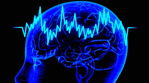
Getty Images Stories involving infidelity, politics, war, labor strikes, or abortion often tap into our most deeply held values. Recent research found that value-laden stories activate the default mode network, previously thought to activate as the brain’s autopilot. Researchers now believe the default mode network is activated as the brain works to find meaning in narrative. The study was discussed in a recent article in MedicalXpress:
Everyone has at least a few non-negotiable values. These are the things that, no matter what the circumstance, you'd never compromise for any reason - such as "I'd never hurt a child," or "I'm against the death penalty."
Real-time brain scans show that when people read stories that deal with these core, protected values, the "default mode network" in their brains activates.
This network was once thought of as just the brain's autopilot, since it has been shown to be active when you're not engaged by anything in the outside world - but studies like this one suggest that it's actually working to find meaning in the narratives.
"The brain is devoting a huge amount of energy to whatever that network is doing. We need to understand why," said Jonas Kaplan of the USC Dornsife Brain and Creativity Institute. Kaplan was the lead author of the study, which was published on Jan. 7 in the journal Cerebral Cortex.
Kaplan thinks that it's not just that the brain is presented with a moral quandary, but rather that the quandary is presented in a narrative format.
"Stories help us to organize information in a unique way," he said.
To find relevant stories, the researchers sorted through 20 million blog posts using software developed at the USC Institute for Creative Technologies.
"We wanted to know how people tell stories in their daily lives. It was kind of like finding stories in their natural habitat," said Kaplan, assistant research professor of psychology at the Brain and Creativity Institute at the USC Dornsife College of Letters, Arts and Sciences.
That 20 million was pared down to 40 stories that each contained an example of a crisis involving a potentially protected value: cheating on a spouse, having an abortion, crossing a picket line, or getting in a fight.
Those stories were translated into Mandarin Chinese and Farsi, and then read by American, Chinese and Iranian participants in their native language while their brains were scanned by fMRI. They also answered general questions about the stories while being scanned.
Stories that participants said involved values that were protected to them activated the default mode network in their brain to a greater degree. In addition, the level of activation varied from culture to culture. On average, Iranians showed the greatest level of activation in the study, while the Chinese participants showed the least.
"Stories appear to be a fundamental way in which the brain organizes information in a practical and memorable manner. It is important to understand the neural mechanisms required to do this, and this study is a step in that direction," said Antonio Damasio, senior author of the study. Damasio is co-director of the Brain and Creativity Institute, holder of the David Dornsife Chair in Neuroscience and a professor of psychology and neurology.
It's not yet clear whether a value either is or is not protected, or whether the sacredness of a value is on a sliding scale. But in a nation where political beliefs are growing more polarized and entrenched, it's important to understand what biological processes lie at the root of these values, Kaplan said.
"People will often hold political values as protected values and protected values are at the root of many political conflicts around the world, which is why they're interesting to us," he said.
Read the original article Here
- Comments (0)
- Brainwaves Spread Through Mild Electrical Field
- By Jason von Stietz, M.A.
- January 15, 2016
-
iStockPhoto Scientists once thought that the brain’s electrical endogenous fields were too weak to propagate transmission. However, recent findings from researchers from Case Western Reserve University suggested that brainwaves spread through a mild electrical field. The study was discussed in a recent article in MedicalXpress:
Researchers at Case Western Reserve University may have found a new way information is communicated throughout the brain.
Their discovery could lead to identifying possible new targets to investigate brain waves associated with memory and epilepsy and better understand healthy physiology.
They recorded neural spikes traveling at a speed too slow for known mechanisms to circulate throughout the brain. The only explanation, the scientists say, is the wave is spread by a mild electrical field they could detect. Computer modeling and in-vitro testing support their theory.
"Others have been working on such phenomena for decades, but no one has ever made these connections," said Steven J. Schiff, director of the Center for Neural Engineering at Penn State University, who was not involved in the study. "The implications are that such directed fields can be used to modulate both pathological activities, such as seizures, and to interact with cognitive rhythms that help regulate a variety of processes in the brain."
Scientists Dominique Durand, Elmer Lincoln Lindseth Professor in Biomedical Engineering at Case School of Engineering and leader of the research, former graduate student Chen Sui and current PhD students Rajat Shivacharan and Mingming Zhang, report their findings in The Journal of Neuroscience.
"Researchers have thought that the brain's endogenous electrical fields are too weak to propagate wave transmission," Durand said. "But it appears the brain may be using the fields to communicate without synaptic transmissions, gap junctions or diffusion."
How the fields may work
Computer modeling and testing on mouse hippocampi (the central part of the brain associated with memory and spatial navigation) in the lab indicate the field begins in one cell or group of cells.
Although the electrical field is of low amplitude, the field excites and activates immediate neighbors, which, in turn, excite and activate immediate neighbors, and so on across the brain at a rate of about 0.1 meter per second.
Blocking the endogenous electrical field in the mouse hippocampus and increasing the distance between cells in the computer model and in-vitro both slowed the speed of the wave.
These results, the researchers say, confirm that the propagation mechanism for the activity is consistent with the electrical field.
Because sleep waves and theta waves—which are associated with forming memories during sleep—and epileptic seizure waves travel at about 1 meter per second, the researchers are now investigating whether the electrical fields play a role in normal physiology and in epilepsy.
If so, they will try to discern what information the fields may be carrying. Durand's lab is also investigating where the endogenous spikes come from.
Read the original article Here
- Comments (1)
- Prosocial Behavior Linked to Amygdala
- By Jason von Stietz, M.A.
- December 30, 2015
-

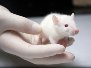
Getty Images Researchers studying social behavior of rhesus monkeys found amgydala activation linked to prosocial or charitable behavior. Findings suggested that when rhesus monkeys were faced with a task of either donating or withholding rewards to others, activation of the amgydala reflected the value of the rewards delivered. The study was discussed in a recent article in MedicalXpress:
The amygdala, a small structure at the front end of the brain's temporal lobe, has long been associated with negative behaviors generally, and specifically with fear. But new research from Michael Platt, the James S. Riepe University Professor in the psychology, neuroscience and marketing departments at the University of Pennsylvania, along with Steve Chang from Yale University and collaborators from Duke, shows this collection of nuclei can also influence positive social functions like kindness and what might be called charitable giving in humans.
Such a link could have implications for people with autism, schizophrenia or anxiety-related disorders, Platt said.
"What we're trying to do is both identify and understand the basic brain mechanism that allows us to be kind to each other and to respond to the experiences of other individuals," he said. "We're also trying to use that knowledge to evaluate potential therapies that could improve the function of these neural circuits, especially for those who have difficulty connecting with others."
To make this discovery about the amygdala, Platt and his team looked at the social behavior of rhesus macaques, a non-human primate species he has studied for 22 years both in the lab and in the wild, on an island off of Puerto Rico called Cayo Santiago. The researchers incorporated a task they developed four years ago as a way to observe how animals make beneficial decisions, a process Platt described as a reward-donation task.
"We have an actor monkey and a recipient monkey. The actor monkey learns that different-colored shapes on the screen are associated with a reward that can be delivered to himself, to the monkey next to him, to both or to nobody at all," he said. "They learn that over a couple of weeks."
Once the researchers determine the monkeys understand the task, based on how quickly the macaques respond to the rewards, they then present the actor monkey with choices and their accompanying potential rewards. The primates can keep the reward (in this case, a squirt of juice), share it, give it away or let it go to waste.
"Generally our actor monkeys prefer to reward the other monkey rather than let it go unclaimed," Platt said. Relationship status matters, too. "They are more likely to give to those they're more familiar with," he added, "and also to monkeys subordinate to them. The social relationships shape how prosocial the actor monkeys are."
Simultaneous to watching the monkeys' behavior, Platt and his colleagues recorded the neural activity of the amygdala of each animal, to note any correlations between what's happening in the brain and their outward actions. They found that neural activity in the amygdala reflected the value of the recipient's reward in the same way it reflected the value of the reward for the actor. The scientists could predict when actors would give rewards to the recipients based on these neural responses.
When oxytocin was introduced, behaviors changed rapidly. Oxytocin is a hormone linked to social bonds between individuals. In animals, it's been shown to create strong ties between mother and offspring, as well as male and female partners in certain monogamous animals. Though not definitive, research on how oxytocin affects humans has, in some cases, shown to help those with autism better read and understand social cues.
"We don't really know how it works in people. It's very difficult to study. When people inhale oxytocin, there is a change in blood flow to the amygdala, which we think might be involved in making people kinder and more receptive to others," Platt said. In his experiment, the monkeys receiving oxytocin became more willing to give to other monkeys and paid more attention to them after offering the rewards.
Rhesus macaques offer a valuable comparison to humans because the animals model many of the social behaviors in which humans engage. They also live in large social groups and form what Platt described as long-term social bonds.
"Not only with relatives but non-relatives, too. You can think of these as friendships or alliances, and they work hard to maintain them," he said. "Just like humans, the stronger these bonds the monkeys have, the more successful they are. Monkeys with more friends and better friends live longer and have more offspring."
Thanks to funding from the Simons Foundation, the next phase of this research will focus on the part of the brain that identifies social context, for example, determining who is nearby, for what reason and what effect each decision will have on them.
Read the original article Here
- Comments (0)
- Social Decision-Making in Autism
- By Jason von Stietz, M.A.
- December 23, 2015
-
Getty Images It is widely believed that individuals with autism lack the capacity to read social cues. However, recent findings suggest autism affects not how social information is processed but how the information is used in decision making. The study was discussed in a recent article in MedicalXpress:
Scientists at the Max Planck Institute of Psychiatry in Munich together with colleagues in Cologne and Zürich have used mathematical models to explain differences in social behaviour associated with autistic personality traits. They show that autistic traits do not – as previously thought – stop the individual from "reading" social cues, but instead affect how social information is used in making decisions. This new understanding provides a new basis for future research that will improve therapies for people with autism.
Autism spectrum disorder is a common psychiatric disorder that is characterized from childhood onwards by major impairments in social interaction and communication. About one half of autistic individuals are "high-functioning" as they have no intellectual impairment. However, all are severely limited in their social and professional life, which is why so many people with autism also suffer from depression. It has been suggested that autistic individuals may not "read" social cues and as a result do not respond to them properly. Alternatively, it has been suggested they are unable to empathize, thereby leading to social problems.
Leonhard Schilbach, consultant psychiatrist and Research Group leader at the Max Planck Institute of Psychiatry, explains "we have shown in this study that autistic trait-related differences in social behaviour are not due to a general inability to process social cues, but are related to the way social information is taken into account during decision-making".
It is thought that reading cues and making decisions involve an interplay between "expectations" generated in higher brain areas and signals from lower sensory brain areas. Normally, the behaviour of others that causes the sensory signal is interpreted in comparison to such "expectations". This allows individuals to regulate their behaviour in a context-sensitive and socially adequate way. In contrast to this, autism might be the result of an imbalance in this interplay, with a stronger reliance on sensory signals.
Computational modeling uses mathematical descriptions to characterize the variables that underlie social behaviour. Previous research has shown that humans do not only "read" the overt behaviour of others, but also the intentions that underlie and motivate the behaviour and use this information when making. Lead scientist Leonhard Schilbach explains, "we used a computational approach to investigate whether differences in social decision-making observed in people who are high or low in autistic traits are related to an inability to monitor others' intentions".
Schilbach and colleagues found that higher autistic traits do not impair the ability to process social information per se, but rather how this information influences decision-making. Furthermore, it was observed that individuals high in autistic traits found it harder to optimally use social information during unpredictable situations. It is hoped that this new insight into the autism spectrum will provide improved interventions for patients. Furthermore, the computational approach used in this study could also help to characterize social difficulties in other psychiatric disorders.
Read the article Here
- Comments (1)
- Enhancing Experience-Dependent Neuroplasticity
- By Jason von Stietz, M.A.
- December 19, 2015
-
Getty Images Experience-dependent neuroplasticity, the brain’s capacity to change in response to environmental stimuli and learning, is a fundamental property of the brain. The impairment of this function in the brain is related to many psychiatric disorders including depression and bipolar disorder. Researchers recently studied the effect of D-cycloserine on the brain’s capacity for experience-dependent neuroplasticity (measured by EEG). The study was recently discussed in an article in MedicalXpress:
In a recent study published in the Proceedings of the National Academy of Sciences, a group of researchers from various U.S. colleges have collaborated to determine if augmenting the signaling of a particular brain receptor would boost neuroplasticity in adults. During early development, experience-dependent neuroplasticity actually interacts with genetic programming in order to establish the neuronal organization and functionally connected circuits that characterize the mature brain.
This basic circuitry is well established by early adulthood, but throughout the lifespan, adult brains depend on experience-dependent neuroplasticity to enable new behavior patterns. Given the general acceptance of the relatively new idea that neuroplasticity endures in adults, the ability to augment its mechanisms could yield new approaches to associated psychiatric disorders. Here, the researchers sought to determine if augmenting N-methyl-D-aspartate receptor (NMDAR) signaling would promote experience-dependent plasticity. They tested a drug called D-cycloserine (DCS) on a group of participants who were monitored via a recently developed EEG paradigm for changes in plasticity.
The participants, divided into groups that received either DCS or placebo, engaged in three cognitive tasks: A weather prediction task, an information integration task and an n-back task, once before administration of DCS or placebo, and again 31 hours later. They determined that participants who received DCS showed greater potentiation of plasticity following the high-frequency visual stimuli of the tests than did those who received placebo. "Our findings of enhanced acquisition of the weather prediction task and the information integration task are consistent with other findings of enhanced incremental learning following DCS administration, including on category learning, motor learning, and mental rotation learning tasks," the authors write.
They note that the performance of DCS participants on the n-back test, which was a spatial working memorytask, did not differ measurably from the performance of those receiving placebo. They note that this result is consistent with a growing body of evidence that the transient memory underlying working memory is modulated in a fundamentally different way than experience-dependent neuroplasticity.
While noting the limitation that the study was restricted to healthy young adults, the authors conclude that their results strongly suggest that enhancing NMDAR signaling augments experience-dependent plasticity in adult brains across a variety of tasks that leverage that ability. "These findings suggest exciting possibilities for using NMDAR agonists to help ameliorate plasticity deficits in neurodegenerative and psychiatric disorders. Our results complement a growing literature that suggests that DCS can enhance new learning during cognitive behavioral therapy interventions and cognitive training programs."
The researchers suggest that parallel studies in older adults and patient groups are an obligatory next step in assessing DCS as a therapeutic intervention for psychiatric disorders.
Read the full article Here
- Comments (0)
- Surgical Treatment of TBI in Older Adults
- By Jason von Stietz, M.A.
- November 30, 2015
-

Getty Images Researches at the Helsinki University Hospital Department of Neurosurgery investigated these use of surgery to treat acute subdural hemotomas in patients over the age of 75. Previously, older adult patients were not treated surgically, as the rates of patents who survived and recovered successfully were low. However, new findings show that older adult patients who lived independently before the accident, were not taking anticoagulants, and who arrived at the hospital conscious received were treated successfully through surgery. The study was discussed in a recent article in NeuroScientistNews:
According to a study completed at the Helsinki University Hospital Department of Neurosurgery, even patients over the age of 75 may recover from severe traumatic brain injury. This is the first study to describe the results of surgically treated elderly patients with acute subdural hematomas.
It is generally accepted that elderly patients who suffer from an acute subdural hematoma should not be treated surgically, as few survive and even fewer recover to an independent life. However, the world's population is rapidly aging leading to an increased rate of fall accidents. In the worst case, falling may result in brain hemorrhage.
Age is one of the most significant outcome predictors in patients with traumatic brain injury. If the patient is young, an acute subdural hematoma is normally treated through a neurosurgical operation. However, even among young patients, mortality and significant morbidity are highly common, despite surgical treatment. In older patients, the success rate of the surgery are made worse by the fact that many patients are typically using oral anticoagulant medications to treat other cardiovascular diseases.
The Neurosurgical Department in Helsinki University Hospital has been an exception in its policy to also treat elderly patients with acute subdural hematomas surgically. Researchers from the University of Helsinki and Helsinki University Hospital have now determined how the patients' functional status before the injury and the use of oral anticoagulant medications influence the prognosis of patients 75 years or older operated on for an acute subdural hematoma.
The study showed that no patients who had been brought to hospital unconscious, who had not been independent before the trauma, or who had used anticoagulants were alive at one year after the surgery.
"What was surprising, however, was that patients who were conscious at presentation, who were not using anticoagulants or were independent before the operation, recovered quite well. The expected lifespan of these patients was comparable to their age-matched peers," says MD, PhD Rahul Raj, one of the main authors.
"One should be careful to make to strong conclusions from such a small number of patients," Raj points out, "but it seems that in approximately half of all cases, even elderly patients may benefit from surgery and recover to an independent life. It is important to note that included patients had an isolated acute subdural hematoma with no injuries to the brain tissue itself. This means that the results cannot be applied to patients with contusions or other intracranial injuries, whose treatment and prognosis are different."
The decision to operate should not be based on age alone
According to Raj, the study throws new light on the old assumption that surgical treatment of the elderly is not a sensible course of action: "The decision to treat through surgery should not be based on age alone, even though this is common."
Surgery of an acute subdural hematoma followed by intensive care and rehabilitation involve major costs and can cause significant suffering to patients and relatives. Thus, it is important to perform surgery on only the patients who are likely to benefit from it.
"But how do you define a bad prognosis? If only one in ten patients recovers sufficiently to live at home, is the treatment worthwhile? If half of the treated patients die within the year, is the treatment worthwhile? This is not a medical decision," the researchers emphasize. They believe that in the future, surgical treatment will be increasingly restricted to patients with the highest likelihood of recovering.
Read the original article Here
- Comments (2)
- Brain's Stress Circuitry Involved in Alzheimer's Disease Treatment
- By Jason von Stietz, M.A.
- November 27, 2015
-
Getty Images Researchers at the University of San Diego School of Medicine investigated the use of a small molecule drug in preventing and treating Alzheimer’s disease in mice. Researchers found that the drug significantly reduces activity of the stress circuitry in the brains of the mice and resulted in the prevention of neurodegeneration and cognitive impairment. The study was discussed in a recent article of NeuroScientistNews:
The findings are described in the current online issue of the journal Alzheimer’s & Dementia: The Journal of the Alzheimer’s Association.
The results underscore the complexity and diversity of AD, whose causes appear to be a mix of genetic, lifestyle and environmental factors. Previous research has shown a link between the brain’s stress signaling pathways and AD. Specifically, the release of a stress-coping hormone called corticotropin-releasing factor (CRF), which is widely found in the brain and acts as a neurotransmitter/neuromodulator, is dysregulated in AD and is associated with impaired cognition and with detrimental changes in tau protein and increased production of amyloid-beta—protein fragments that clump together and trigger the neurodegeneration characteristic of AD.
“Our work and that of our colleagues on stress and CRF have been mechanistically implicated in Alzheimer’s disease, but agents that impact CRF signaling have not been carefully tested for therapeutic efficacy or long-term safety in animal models,” said the study’s principal investigator and corresponding author Robert Rissman, PhD, assistant professor in the Department of Neurosciences and Biomarker Core Director for the Alzheimer’s Disease Cooperative Study (ADCS).
“The novelty of this study is two-fold: We used a preclinical prevention paradigm of a CRF-antagonist (a drug that blocks the CRF receptor in brain cells) called R121919 in a well-established AD model – and we did so in a way that draws upon our experience in human trials. We found that R121919 antagonism of CRF-receptor-1 prevented onset of cognitive impairment and synaptic/dendritic loss in AD mice.”
In other words, the researchers determined that modulating the mouse brain’s stress circuitry (without actually changing the normal response) mitigated generation and accumulation of amyloid plaques widely attributed with causing neuronal damage and death. As a consequence, behavioral indicators of AD were prevented and cellular damage was reduced. The mice began treatment at 30-days-old – before any pathological or cognitive signs of AD were present – and continued until six months of age.
One particular challenge, Rissman noted, is limiting exposure of the drug to the brain so that it does not impact the body’s ability to response to stress. “This can be accomplished because one advantage of these types of small molecule drugs is that they readily cross the blood-brain barrier and actually prefer to act in the brain,” Rissman said. Drugs like R121919 were originally designed to treat generalized anxiety disorder, irritable bowel syndrome and other diseases, but failed to be effective in treating those disorders.
“Rissman’s prior work demonstrated that CRF and its receptors are integrally involved in changes in another AD hallmark, tau phosphorylation,” said William Mobley, MD, PhD, chair of the Department of Neurosciences and interim co-director of the Alzheimer’s Disease Cooperative Study at UC San Diego. “This new study extends those original mechanistic findings to the amyloid pathway and preservation of cellular and synaptic connections. Work like this is an excellent example of UC San Diego’s bench-to-bedside legacy, whereby we can quickly move our basic science findings into the clinic for testing,” said Mobley.
Rissman said R121919 was well-tolerated by AD mice (no significant adverse effects) and deemed safe, suggesting CRF-antagonism is a viable, disease-modifying therapy for AD. Rissman noted that repurposing R121919 for human use was likely not possible at this point. He and colleagues are collaborating with the Sanford Burnham Prebys Medical Discovery Institute to design new assays to discover the next generation of CRF receptor-1 antagonists for testing in early phase human safety trials.
“More work remains to be done, but this is the kind of basic research that is fundamental to ultimately finding a way to cure – or even prevent – Alzheimer’s disease,” said David Brenner, MD, vice chancellor, UC San Diego Health Sciences and dean of UC San Diego School of Medicine. “These findings by Dr. Rissman and his colleagues at UC San Diego and at collaborating institutions on the Mesa suggest we are on the cusp of creating truly effective therapies.”
Read the full article Here
- Comments (3)
- The Brain Conserves Energy By Forgetting
- By Jason von Stietz, M.A.
- November 11, 2015
-
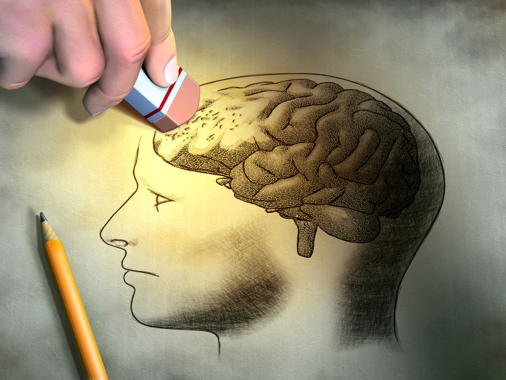
Photo Credit: Shutterstock How is it that we seem to forget information that we deem useless or unnecessary? Researchers at the Lund University in Sweden have investigated one of the brain’s mechanisms of forgetting or ignoring such information. The study was discussed in a recent article by MedicalXpress:
Our brains not only contain learning mechanisms but also forgetting mechanisms that erase "unnecessary" learning. A research group at Lund University in Sweden has now been able to describe one of these mechanisms at the cellular level.
The group's results, published in the international journal Proceedings of the National Academy of Sciences of the United States of America (PNAS), explain a theoretical learning phenomenon which has so far been difficult to understand.
The premise is that human or animal subjects can learn to associate a certain tone or light signal with a puff of air to the eye. The air puff makes the subject blink, and eventually they blink as soon as they hear the tone or see the light signal. The strange thing, however, is that if the tone and the light are presented together (and with the air puff), the learning does not improve, but gets worse.
"Two stimuli therfore achieve worse results than just one. It seems contrary to common sense, but we believe that the reason for it is that the brain wants to save energy", says brain researcher and professor Germund Hesslow.
His colleague Anders Rasmussen, who performed the present study, has previously shown that when the brain has learnt a particular association sufficiently, certain neurons that act as a brake on the learning mechanism, are activated.
"You could say that the part of the brain that learned the association (a part of the brain called the cerebellum) is telling its 'teacher': 'I know this now, please be quiet'. When the brain has learnt two associations, the brake becomes much more powerful. That is why it results in forgetting, usually only temporarily, however", explains Germund Hesslow.
Maintaining unnecessary association pathways requires energy for the brain. The researchers believe that this is the reason for the brake mechanism – even though in this case it happened to be a little too powerful.
The Lund researchers were able to describe how the nerve cells learn and forget through studies of animals, but believe that the mechanisms are likely to be the same in the human brain. Therefore, these findings are of fundamental interest for both brain researchers and psychologists. They could also be of practical interest to educators.
"Obviously, it should be important for teachers to know the mechanisms by which the brain erases the things it considers unnecessary. You do not want to accidentally activate these mechanisms", says Germund Hesslow.
Read the orginal article Here
- Comments (0)
- ADHD Improperly Diagnosed in Children
- By Jason von Stietz, M.A.
- October 30, 2015
-

Getty Images What is causing the rise in children being diagnosed with ADHD? Is it that the modern educational environment requires more of children, which makes it harder for ADHD to go unnoticed? Is it the exposure to chemicals in our environment or the processed sugar in our diet. A study by the Center for Disease Control and Prevention found that ADHD is often improperly diagnosed. The findings were discussed in an article by the Washington Post:
All sorts of theories have been proposed to explain the alarming rise -- 6.4 million in 2011, a 42 percent jump from 2004 -- in schoolchildren being diagnosed with Attention-Deficit/Hyperactivity Disorder, or ADHD, requiring therapy, medicine or both to make it through their day.
Some believe it's simply a matter of more awareness (and paranoia) -- meaning that more parents are seeking a diagnosis. Others wonder if it's schools (they're more academic now than in the past, requiring kids to sit still for longer periods of time making those who have ADHD more obvious).
Still others blame the environment (all those chemicals we use). Or diet (yet another thing to blame on processed sugar).
The CDC report takes an in-depth look at how children with ADHD came to get the label through a survey of 2,976 families. While in the majority of cases health care providers followed American Academy of Pediatrics guidelines when making a diagnosis, there was still a large number of children for whom these practices weren't followed.
In 18 percent of cases, the diagnosis was done solely on the basis of family members' reports, which is inconsistent with AAP recommendations that information be collected from individuals across multiple settings -- such as a teacher, piano instructor, or sports coach. Additionally, one out of every 10 children was diagnosed without the use of a behavior rating scale that is supposed to be administered.
The study also shows that children are getting diagnosed at an earlier age, with half being diagnosed at age 6 or below: 17.1 percent at age 6, 14.6 percent at age 5, and 16 percent at age 4 or younger.
Read the original article Here
- Comments (0)
- High Connectivity Related to Positive Traits
- By Jason von Stietz, M.A.
- October 17, 2015
-
Getty Images Researchers involved in the Human Connectome Project made a surprising discovery when examining the relationship between brain connectivity during a resting state and several factors such as education, socioeconomic status, substance use, and personality traits. It was found that, overall, positive traits related to increased connectivity whereas negative traits related to decreased connectivity. Those with more highly connected brains were more likely to be more highly educated and perform better on memory tests. Those with less brain connectivity were more likely to smoke and have more aggressive personalities. The study and it’s implications were discussed in a recent article in Scientific American:
The brain’s wiring patterns can shed light on a person’s positive and negative traits, researchers report in Nature Neuroscience. The finding, published on September 28, is the first from the Human Connectome Project (HCP), an international effort to map active connections between neurons in different parts of the brain.
The HCP, which launched in 2010 at a cost of US$40 million, seeks to scan the brain networks, or connectomes, of 1,200 adults. Among its goals is to chart the networks that are active when the brain is idle; these are thought to keep the different parts of the brain connected in case they need to perform a task.
In April, a branch of the project led by one of the HCP's co-chairs, biomedical engineer Stephen Smith at the University of Oxford, UK, released a database of resting-state connectomes from about 460 people between 22 and 35 years old. Each brain scan is supplemented by information on approximately 280 traits, such as the person's age, whether they have a history of drug use, their socioeconomic status and personality traits, and their performance on various intelligence tests.
Axis of connectivity
Smith and his colleagues ran a massive computer analysis to look at how these traits varied among the volunteers, and how the traits correlated with different brain connectivity patterns. The team was surprised to find a single, stark difference in the way brains were connected. People with more 'positive' variables, such as more education, better physical endurance and above-average performance on memory tests, shared the same patterns. Their brains seemed to be more strongly connected than those of people with 'negative' traits such as smoking, aggressive behaviour or a family history of alcohol abuse.Marcus Raichle, a neuroscientist at Washington University in St Louis, Missouri, is impressed that the activity and anatomy of the brains alone were enough to reveal this 'positive-negative' axis. “You can distinguish people with successful traits and successful lives versus those who are not so successful,” he says.
But Raichle says that it is impossible to determine from this study how different traits relate to one another and whether the weakened brain connections are the cause or effect of negative traits. And although the patterns are clear across the large group of HCP volunteers, it might be some time before these connectivity patterns could be used to predict risks and traits in a given individual. Deanna Barch, a psychologist at Washington University who co-authored the latest study, says that once these causal relationships are better understood, it might be possible to push brains toward the 'good' end of the axis.
Van Wedeen, a neuroscientist at Massachusetts General Hospital in Boston, says that the findings could help to prioritize future research. For instance, one of the negative traits that pulled a brain farthest down the negative axis was marijuana use in recent weeks. Wedeen says that the finding emphasizes the importance of projects such as one launched by the US National Institute on Drug Abuse last week, which will follow 10,000 adolescents for 10 years to determine how marijuana and other drugs affect their brains.
Wedeen finds it interesting that the wiring patterns associated with people's general intelligence scores were not exactly the same as the patterns for individual measures of cognition—people with good hand–eye coordination, for instance, fell farther down the negative axis than did those with good verbal memory. This suggests that the biology underlying cognition might be more complex than our current definition of general intelligence, and that it could be influenced by demographic and behavioural factors. “Maybe it will cause us to reconsider what [the test for general intelligence] is measuring,” he says. “We have a new mystery now.”
Much more connectome data should emerge in the next few years. The Harvard Aging Brain Study, for instance, is measuring active brain connections in 284 people aged between 65 and 90, and released its first data earlier this year. And Smith is running the Developing Human Connectome Project in the United Kingdom, which is imaging the brains of 1,200 babies before and after birth. He expects to release its first data in the next few months. Meanwhile, the HCP is analysing genetic data from its participants, which include a large number of identical and fraternal twins, to determine how genetic and environmental factors relate to brain connectivity patterns.
See the original article Here
- Comments (0)
 Subscribe to our Feed via RSS