Blog
- Study Links Affective Understanding to Attraction
- By Jason von Stietz, M.A.
- April 8, 2016
-

Getty Images What is it that leads to attraction? Researchers conducted an experiment utilizing fMRI scans of participants watching a video of a woman. It was found that participants found her more attractive, which was reflected in the reward center of the brain, when they perceived themselves to understand her emotional experience. The findings were discussed in a recent article in Medical Xpress:
(Medical Xpress)—A team of researchers with members from a large number of institutions in Germany has conducted a study that has revealed more about the way interpersonal attraction works in the brain. In their paper published in Proceedings of the National Academy of Sciences, the group describes two experiments they conducted with volunteers, their results and what they believe was revealed about the nature of the mechanism of attraction between people.
Most everyone has experienced near instant attraction to someone else, whether of a social or sexual nature, but few are able to pin down exactly why they felt that attraction. Based on two experiments they conducted with human volunteers, the researchers suggest it may have to do with matchingneural circuitry.
To learn more about attraction, the researchers ran two experiments, the first consisted of showing 19 male and 21 female volunteers, videos of six different women as they experienced fear or sadness. The volunteers were asked to choose which emotion was being shown, and then to mark down how confident they were in their choice. To gauge how much of an attraction they volunteers felt for the women in the videos, they were asked to enlarge a picture of the woman both before and after seeing her in the video—each was also asked to answer questions about each woman, such as how much they would like to meet her in real life, if she would understand them, etc.
The second experiment was run with a different set of volunteers who were also asked to watch the woman in the videos, but the second group did so while undergoing an fMRI imaging—the researchers were specifically looking for activity in the part of the brain known to be associated with rewards.
The final phase of the experiment involved combining data from both experiments to see if any patterns might emerge. The researchers report that most of the volunteers were able to identify the emotions being portrayed, and the more confident they felt they were able to identify the correct emotion, the more attracted to her they felt. This was confirmed in the fMRI scans—reward centers in the volunteers' brains lit up more when watching women they felt they could read their emotions better.
The researchers propose that their results suggest that in addition to physical attractiveness, people are attracted to other people due to their own feelings of similarity to another person, which gives them a feeling of understanding, or connectedness.
Read the original article Here
- Comments (0)
- Neuronal Feedback Changes Our Perception
- By Jason von Stietz, M.A.
- April 4, 2016
-

Photo Credit: Carnegie Mellon University Have you ever thought you saw one thing, but when you looked again it was something else? Often times the brain notices the beginning of a visual pattern and will automatically connect the rest of the dots. How does this happen? Researchers at the Carnegie Mellon University studied top-down visual processing. Findings were discussed in a recent article in MedicalXpress:
Understanding this feedback system could provide new insight into thevisual system's neuronal circuitry and could have further implications for understanding how the brain interprets and understands sensory stimuli.
Many optical illusions make you see something that's not there. Take the Kanizsa triangle: when you place three Pac-Man-like wedges in the right spot, you see a triangle, even though the edges of the triangle aren't drawn.
"We see with both our brain and our eyes. Your brain is making inferences that allow you to see the triangle. It's connecting the dots between the corners of the wedges," said Kuhlman, who is a member of Carnegie Mellon's BrainHub neuroscience initiative and the joint Carnegie Mellon/University of Pittsburgh Center for the Neural Basis of Cognition (CNBC). "Optical illusions illustrate some of the amazing things our visual system can do."
When we look at an object, information about what we see travels through circuits of neurons beginning in the retina, through the thalamus and into the brain's visual cortex. In the visual cortex, the information gets processed in multiple stages and is ultimately sent to the prefrontal cortex—the area of the brain that makes decisions, including how to respond to a given stimulus.
However, not all information stays on this forward moving path. At the secondary stage of processing in the visual cortex some neurons reverse course and send information back to the first stage of processing. Researchers at Carnegie Mellon wondered if this feedback could change how the neurons in the visual cortex respond to a stimulus and alter the messages being sent to the prefrontal cortex.
While there has been a good deal of research studying how information moves forward through the visual system, less has been done to study the impact of the information that moves backward. To find out if the information traveling from the secondary stage of processing back to the first stage impacted how information is encoded in the visual system, the researchers needed to quantify the magnitude of information that was being sent from the second stage back to the first stage. Using a mouse model, they recorded normal neuronal firing in the first stage of the visual cortex as the mouse looked at moving patterns that represented edges. They then silenced the neurons in the second stage using modified optogenetic technology. This halted the feedback of information from the second stage back to the first stage, and allowed the researchers to determine how much of the neuronal activity in the first stage of visual processing was the result of feedback.
Twenty percent of the neuronal activity in the visual cortex was the result of feedback, a concept Kuhlman calls reciprocal connectivity. This indicates that some of the information coming from the visual cortex is not a direct response to a visual stimuli, but is a response to how the stimuli was perceived by higher cortical areas.
The feedback, she says, might be what causes our brain to complete the undrawn lines in the Kanizsa triangle. But more importantly, it signifies that studying neuronal feedback is important to our understanding of how the brain works to process stimuli.
"This represents a new way to study visual perception and neural computation. If we want to truly understand the visual pathway, and cortical function in general, we have to understand these reciprocal connection," Kuhlman said.
Read the original artice Here
- Comments (0)
- fMRI Utilized in Neurofeedback
- By Jason von Stietz, M.A.
- March 25, 2016
-
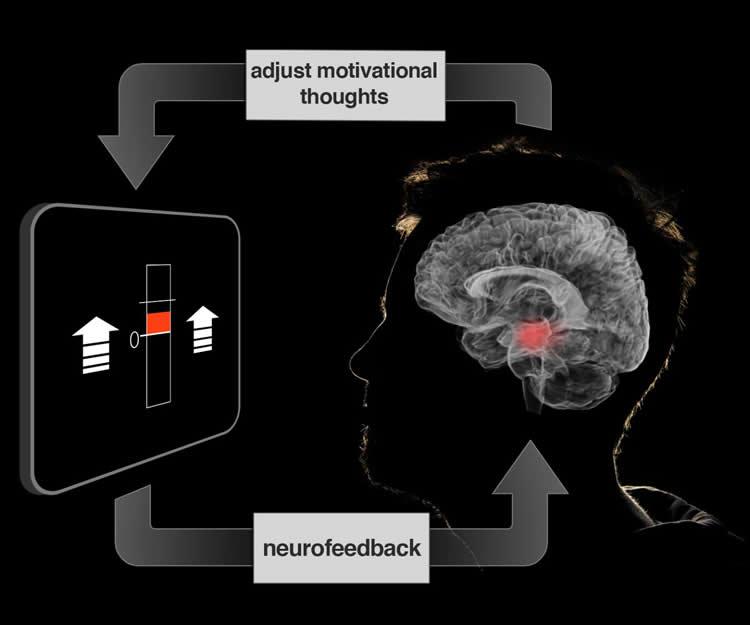
Credit: Jeff MacInnes, Duke University. Although neurofeedback has traditionally relied on the use of EEG, researchers from Duke University have examined the use of fMRI data. Participants’ brain activity was monitored and feedback was provided to them in the form of a fluctuating thermometer. Both EEG and fMRI neurofeedback were discussed in a recent article in NeuroscienceNews:
At our best, we motivate ourselves every day to get dressed and go to work or school. Although there are larger incentives at work, it’s our own volition that powers us through our innumerable daily tasks.
If we could learn to control the motivational centers of our brains that drive volition, would it lead us toward healthier, more productive lives? Using a new brain imaging strategy, Duke University scientists have now taken a first step in understanding how to manipulate specific neural circuits using thoughts and imagery.
The technique, which is described in the March 16 issue of the journal Neuron, is part of a larger approach called ‘neurofeedback,’ which gives participants a dynamic readout of brain activity, in this case from a brain area critical for motivation.
“These methods show a direct route for manipulating brain networks centrally involved in healthy brain function and daily behavior,” said the study’s senior investigator R. Alison Adcock, an assistant professor of psychiatry and behavioral sciences and associate director of the Center for Cognitive Neuroscience in the Duke University Institute for Brain Sciences.
Neurofeedback is a specialized form of biofeedback, a technique that allows people to monitor aspects of their own physiology, such as heart rate and skin temperature. It can help generate strategies to overcome anxiety and stress or to cope with other medical conditions.
Neurofeedback has historically relied on electroencephalography or EEG, in which patterns of electrical activity are monitored noninvasively by electrodes attached to the scalp. But these measures provide only rough estimates of where activity occurs in the brain.
In contrast, the new study employed functional magnetic resonance imaging (fMRI), which measures changes in blood oxygen levels, allowing more precisely localized measurements of brain activity.
Adcock’s team has been working on ways to use thoughts and behavior to tune brain function for the past eight years. In this time, they’ve developed tools allowing them to analyze complex brain imaging data in real time and to display it to participants as neurofeedback while they are in the fMRI scanner.
This study focused on the ventral tegmental area (VTA), a small area deep within the brain that is a major source of dopamine, a neurochemical well known for its role in motivation, experiencing rewards, learning, and memory.
According to Adcock’s previous research, when people are given incentives to remember specific images, an increase in VTA activation before the image appears predicts whether the participants are going to successfully remember the image.
External incentives like money work well to stimulate the VTA, but it was unclear whether people could exercise this area on their own, said co-author Jeff MacInnes, a postdoctoral researcher in Adcock’s lab.
In the new study, the team encouraged participants in the scanner to generate feelings of motivation – using their own personal strategies – during 20-second intervals. They weren’t able to raise their VTA activity consistently on their own.
But when the scientists provided participants with neurofeedback from the VTA, presented in the form of a fluctuating thermometer, participants were able to learn which strategies worked, and ultimately adopt more effective strategies. Compared to control groups, the neurofeedback-trained participants successfully elevated their VTA activity.
Participants reported using a variety of different motivational strategies, from imagining parents or coaches encouraging them, to playing out hypothetical scenarios in which their efforts were rewarded, said co-author Kathryn Dickerson, a postdoctoral researcher in Adcock’s group.
The self-generated boost in VTA activation worked even after the thermometer display was removed. Only the participants who had received accurate neurofeedback were able to consistently raise their VTA levels.
“Because this is the first demonstration of its kind, there is much still to be understood,” Adcock added. “But these tools could offer benefits for everyone, particularly those with depression or attention problems.”
The neurofeedback training also activated other regions involved in learning and experiencing rewards, confirming that, at least in the short term, the brain changes its activity more broadly as a result of neurofeedback, Dickerson said.
Adcock said one caveat of the study is that the team has not tested whether the neurofeedback drove changes in behavior. The group is working on those studies now and also plans to conduct the same study in participants with depression and attention deficit hyperactivity disorder (ADHD).
Read the original article Here
- Comments (0)
- Exercise Boosts Neurotransmitters
- By Jason von Stietz, M.A.
- March 18, 2016
-

Getty Images Why does exercise seem to improve our mood? Depression often results in the depletion of two key neurotransmitters: glutamate and GABA. However, recent findings suggest that vigorous exercise boosts the production of these neurotransmitters in the brain. Researchers at University of California Davis examined the effect of exercise on the brain using MRI scans. The study was discussed in a recent article in NeuroScientistNews:
"Major depressive disorder is often characterized by depleted glutamate and GABA, which return to normal when mental health is restored," said study lead author Richard Maddock, professor in the Department of Psychiatry and Behavioral Sciences. "Our study shows that exercise activates the metabolic pathway that replenishes these neurotransmitters."
The research also helps solve a persistent question about the brain, an energy-intensive organ that consumes a lot of fuel in the form of glucose and other carbohydrates during exercise. What does it do with that extra fuel?
"From a metabolic standpoint, vigorous exercise is the most demanding activity the brain encounters, much more intense than calculus or chess, but nobody knows what happens with all that energy," Maddock said. "Apparently, one of the things it's doing is making more neurotransmitters."
The striking change in how the brain uses fuel during exercise has largely been overlooked in brain health research. While the new findings account for a small part of the brain's energy consumption during exercise, they are an important step toward understanding the complexity of brain metabolism. The research also hints at the negative impact sedentary lifestyles might have on brain function, along with the role the brain might play in athletic endurance.
"It is not clear what causes people to 'hit the wall' or get suddenly fatigued when exercising," Maddock said. "We often think of this point in terms of muscles being depleted of oxygen and energy molecules. But part of it may be that the brain has reached its limit."
To understand how exercise affects the brain, the team studied 38 healthy volunteers. Participants exercised on a stationary bicycle, reaching around 85 percent of their predicted maximum heart rate. To measure glutamate and GABA, the researchers conducted a series of imaging studies using a powerful 3-tesla MRI to detect nuclear magnetic resonance spectra, which can identify several compounds based on the magnetic behavior of hydrogen atoms in molecules.
The researchers measured GABA and glutamate levels in two different parts of the brain immediately before and after three vigorous exercise sessions lasting between eight and 20 minutes, and made similar measurements for a control group that did not exercise. Glutamate or GABA levels increased in the participants who exercised, but not among the non-exercisers. Significant increases were found in the visual cortex, which processes visual information, and the anterior cingulate cortex, which helps regulate heart rate, some cognitive functions and emotion. While these gains trailed off over time, there was some evidence of longer-lasting effects.
"There was a correlation between the resting levels of glutamate in the brain and how much people exercised during the preceding week," Maddock said. "It's preliminary information, but it's very encouraging."
These findings point to the possibility that exercise could be used as an alternative therapy for depression. This could be especially important for patients under age 25, who sometimes have more side effects from selective serotonin reuptake inhibitors (SSRIs), anti-depressant medications that adjust neurotransmitter levels.
For follow-up studies, Maddock and the team hope to test whether a less-intense activity, such as walking, offers similar brain benefits. They would also like to use their exercise-plus-imaging method on a study of patients with depression to determine the types of exercise that offer the greatest benefit.
"We are offering another view on why regular physical activity may be important to prevent or treat depression," Maddock said. "Not every depressed person who exercises will improve, but many will. It's possible that we can help identify the patients who would most benefit from an exercise prescription."
Read the original article Here
- Comments (0)
- Studying Prejudice Using EEG
- By Jason von Stietz, M.A.
- March 5, 2016
-
Getty Images Generally, people hold more positive associations toward members of their “in-group” than of an outgroup. For example, sports fan would be quicker to bring to mind words such as “talented” or “great” when describing their favorite team than when describing other teams. This same process has been found to play out when relating to cultural or ethnic in-groups. People are quicker to bring to mind positive associations when presented with an image of someone from their own cultural background. The literature has suggested that this process is involved in unconscious prejudice in which individuals are slower to relate positive attributes to people who are different from them. Researchers from University of Bern investigated this process using EEG. The study was discussed in a recent article in NeuroscienceNews:
We do not always say what we think: we like to hide certain prejudices, sometimes even from ourselves. But unconscious prejudices become visible with tests, because we need a longer time if we must associate unpleasant things with positive terms. Researchers in Bern now show that additional processes in the brain are not responsible for this, but some of them simply take longer.
A soccer fan needs more time to associate a positive word with an opposing club than with his own team. And supporters of a political party associate a favourable attribute faster with their party than with political rivals – even if they endeavour towards the opposite. It is long since known that a positive association with one’s own group, an “in-group”, happens unconsciously faster than with an “outgroup”. These different reaction times become visible in the Implicit Association Test (IAT) with which psychologists examine unconscious processes and prejudices. But why the effort to address a friendly word to an outgroup takes more time was not clear up to now.
Now a team headed by Prof. Daria Knoch from the Department of Social Psychology and SocialNeuroscience at the Institute of Psychology, University of Bern, shows that an additional mental process is not responsible for this, as has often been postulated – but rather the brain lingers longer in certain processes. The study has now been published in the scientific journal PNAS.
Number and sequence of processes are exactly the same
The researchers relied on a unique combination of methods for their study: they conducted an Implicit Association Test with 83 test subjects who are soccer fans or political supporters. While the test persons had to associate positive terms on the screen by means of a button click, either with their in-group or with an outgroup, the brain activity was recorded by means of an EEG (electroen- cephalogram). “We analysed these data with a so-called “microstate analysis”. It enabled us to depict all processes in the brain for the first time – from the presentation of a word up to pressing the button – temporally and also spatially”, explains co-lead author Dr. Lorena Gianotti from the Department of Social Psychology and Social Neuroscience.
The analysis shows the following: the brain runs through seven processes, from the presentation of stimulus – i.e. a word – up to button click, in less than one second. “The number and sequences of these processes remain exactly the same, regardless of whether the test subject had to associate positive words with the in-group, i.e. their club or their party, or with an outgroup”, explains co-lead author Dr. Bastian Schiller, who is in the meantime conducting research at the University of Freiburg.
The reaction time with the outgroup situation is therefore longer, because some of the seven processes take longer – and not because a new process is switched in between. “As a result, corresponding theories can be refuted”, says Schiller. A complete consideration of all processes in the brain is essential for an interpretation, emphasises Lorena Gianotti, and she illustrated this in the following example: on Monday after work you go out to eat with a friend and go to sleep afterwards at 10 pm. On Friday you do exactly the same thing – but you come home two hours later since you can sleep late on the next day. If you now compare the days at 8 pm, both times you were in a restaurant and one could conclude that this is an identical time schedule. If the comparison takes place at 11 pm, you are one time already in bed and one time still on the go. One could think that on Friday you were perhaps still in the sports studio or had an entirely different daily schedule. Therefore it is clear that selective considerations do not allow any conclusion with regard to the entire day – neither with regard to the sequence nor the activities.
“In the research of human behaviour it is essential to consider the underlying brain mechanisms. And this in turn requires suitable methods in order to gain comprehensive findings”, summarises study leader Daria Knoch. A combination of neuroscientific and psychological methods can lead to new insights.
Read the original article Here
- Comments (0)
- Rest is Critical Following a Concussion
- By Jason von Stietz, M.A.
- February 27, 2016
-

Photo Credit: Unknown Researchers at Georgetown University Medical Center (GUMC) found that resting after a concussion is of critical importance to restoring brain health. Using an animal model, researchers investigated the impact of weekly versus daily mild traumatic brain injuries in mice. Findings indicated that neuronal connections lost after an injury were recovered within three days. However, when insults to the brain were daily the neuronal connections were not restored and the impact of the injuries were detected in 1-year follow-up. The study was discussed in a recent article in a recent GUMC press release:
Doctors who order several days of rest after a person suffers a concussion are giving sound advice, say researchers, and new data from animal models explains why.
Georgetown University Medical Center neuroscientists say rest — for more than a day — is critical for allowing the brain to reset neural networks and repair any short-term injury. The new study in mice also shows that repeated mild concussions with only a day to recover between injuries leads to mounting damage and brain inflammation that remains evident a year after injury.
“It is good news that the brain can recover from a hit if given enough time to rest and recover. But on the flip side, we find that the brain does not undertake this rebalancing when impacts come too close together,” says the study’s lead researcher, Mark P. Burns, PhD, assistant professor of neuroscience at GUMC and director of the Laboratory for Brain Injury and Dementia.
This first-of-its-kind study, published in the March 2016 issue of American Journal of Pathology, modeled repeated mild head trauma as a means to investigate brain damage that occurs after a sports, military or domestic abuse injury.
Investigators developed a mouse model of repetitive, extremely mild concussive impacts conducted while the mouse is anesthetized. They compared the brain’s response to a single concussion with an injury received daily for 30 days and one received weekly over 30 weeks.
Mice with a single insult temporarily lose 10-15 percent of the neuronal connections in their brains, but no inflammation or cell death resulted, Burns says. With three days rest, all neuronal connections were restored. This neuronal response is not seen in mice with daily concussions, but the pattern is restored when a week of rest is given between each insult, Burns says.
When a mild concussion occurred each day for a month, inflammation and damage to the brain’s white matter resulted. “This damage became progressively worse for two months and remained apparent one year after the last impact,” Burns says.
“The findings mirror what has been observed about such damage in humans years after a brain injury, especially among athletes,” Burns says. “Studies have shown that almost all people with single concussions spontaneously recover, but athletes who play contact sports are much more susceptible to lasting brain damage. These findings help fill in the picture of how and when concussions and mild head trauma can lead to sustained brain damage.”
Georgetown co-authors are first author Charisse N. Winston, PhD, Maia Parsadanian, David N. Zapple, Sonia Villapol, PhD, and undergraduate students David Barton, Tiffany E. Wilkins, Aidan Neustadtl, Deepa Chellappa, and Andrew D. Alikhani. Contributors also include Emmanuel Planel, PhD, and Anastasia Noel, PhD, from the Centre Hospitalier de l'Université Laval, Neurosciences, Québec, Canada.
The study was supported by Georgetown University’s Neural Injury and Plasticity Training Program, the National Institute for Neurological Disorders and Stroke (R01 NS067417), a supplement to Promote Diversity in Health-Related Research, a donation from KPB Corporation, the Canadian Institute of Health Research, Fonds de Recherche en Santé du Québec and the Natural Sciences and Engineering Research Council of Canada.
Read the oringinal article Here
- Comments (0)
- How Trauma Impacts Memory
- By Jason von Stietz, M.A.
- February 20, 2016
-
Getty Images Some events we remember as vividly as the day they happened. Most people remember every detail about the moment they heard about the events of September 11th 2001. However, how accurate are these vivid and detailed memories? According to recent recent research, not very. This phenomenon was discussed in a recent article of CBCNews:
Where were you, on the morning of Sept. 11, 2001, when you learned that a passenger jet had slammed into the North Tower of the World Trade Center? What were you doing? Who were you with?
Memories of that day, which was deeply painful for so many people, might seem indelibly carved into your mind. There's a good chance, however, you retained far fewer details than you think.
Strongly negative or traumatic experiences are processed and encoded through a distinct neural pathway that filters out "peripheral details," says University of Waterloo cognitive psychologist Myra Fernandes.
The more fear or stress that's generated, the more amplified the filtering effect becomes, especially during experiences that threaten our survival.
The brain locks "on the central aspects of the experience at the expense of the peripheral details," she explains. "The gist is maintained but many of the details are lost.
"From an evolutionary point of view, this is totally plausible," because it emphasizes the parts of a recollection that might serve as a warning later, helping us avoid highly stressful situations in the first place.
'The essential details'
The phenomenon is seen specifically in episodic memories — those of lived experiences that are tied to emotions and context.
The reliability of episodic memory took a central role last week in the ongoing criminal trial of former CBC radio host Jian Ghomeshi, who has pleaded not guilty to four counts of sexual assault and one count of overcoming resistance by choking. The charges involve three women.
It's a common tactic for lawyers to dial in on the minute details of a witness's testimony, pressing to expose any possible contradictions in various retellings of the same story and therefore, the theory goes, raise doubts about its truthfulness.
"By asking someone to repeat a story over and over again, essentially you start to see the story unravel," criminal defence lawyer Daniel Brown explained in an interview with CBC's Metro Morning last week.
Those who say they have lived through trauma, however, are sometimes unable to articulate a coherent narrative owing to the brain's tendency to zero-in on only the most essential elements of what happened.
"The person is overcome with all these essential details," says Lori Haskell, a clinical psychologist in private practice and assistant professor in psychiatry at the University of Toronto. Her research partly focuses on how sexual violence affects the neurobiology of survivors.
"So later on when they try to start creating a narrative, those details aren't accurate – they're not recalled with great detail," she adds.
'Durable' memories
Advanced-imaging studies have shown that traumatic experiences cause two relatively tiny areas deep within the brain, called the amygdala, to kick into overdrive. When the amygdala ramp up, there is a cascade effect through the brain.
Elizabeth Kensinger, a cognitive neuroscientist at Boston College, studies how emotion plays into memory formation.
"The amygdala is activated, and that actually affects how the other regions that take part in processing memory are brought online and how they communicate with one another," she says.
Kensinger's research suggests that while extraneous details may be forgotten, the core components may be less susceptible to the various ways time can erode our recollections.
"There's some really interesting evidence that emotional memories tend to be more durable."
A game of 'telephone'
In general, our episodic memories are "prone to distortions" because they are, in essence, a "reconstruction" of events assembled from building blocks stored throughout the brain. The more we recall any single thing, the greater the chance becomes that we'll remove, or even insert, a block that's not supposed to be there.
Fernandes uses the analogy of the classic children's game "telephone."
"When we recall a memory, we relay it down through our brains … Every time the message is passed on, it changes slightly."
A variety of influences can increase the probability that a recollection will contain erroneous bits. Decades of research by renowned American cognitive psychologist Elizabeth Loftus, for example, has shown that simple, well-crafted linguistic prompts can easily lead someone to unknowingly insert or omit false details into the retelling of a story.
That's not to say all memories contain inaccuracies. In fact, generally speaking, the human brain does an extraordinary job of encoding countless experiences every day.
But it would be too overwhelming to retain all of the information we take in throughout our lives. Research suggests that while we sleep, our brains whittle down experiences — not just traumatic ones — into their most useful parts to make more room, like freeing-up space on a hard drive.
"It's beneficial to our emotional well-being to remember the gist rather than every detail," says Fernandes.
Are you sure?
So, how well do you really remember the details of your day on Sept. 11?
When doing research for The Invisible Gorilla — a book exploring the fallibility of the human mind — University of Illinois cognitive psychologist Daniel Simons wrote down his own answers.
He discovered two of the three people he thought he was with on that morning weren't with him at all, and he has no recollection of being with the person who was there. He's not alone.
More than a dozen universities participated in a survey that asked 2,100 Americans from across the U.S. about their memories of Sept. 11, one, three and 10 years after the attacks.
When all was said and done, 40 per cent of participants told stories notably different than the one that emerged from their original answers.
Interestingly as time passed, those whose answers changed significantly did not become less confident about the accuracy of their stories.
The study is part of a huge body of evidence pointing to the reality that memory is malleable, vulnerable to the curious nature of our own neurobiology.
That doesn't mean we should distrust it, says Simons, but rather, we should appreciate its limits.
Read the original article Here
- Comments (0)
- The Frequencies of Top-Down and Bottom-Up Visual Processing
- By Jason von Stietz, M.A.
- February 12, 2016
-
Getty Images Recent research suggests that the visual cortex processes information in two ways: top-down and bottom-up. Utilizing magnetoencephalography, researchers found that bottom-up information flowed through gamma waves whereas top-down information flowed through alpha-beta waves. The study was discussed in a recent article in NeuroScientistNews:
In the brain, the visual cortex processes visual information and passes it from lower to higher areas of the brain. However, information also flows in the opposite direction, e.g. to direct attention to particular stimuli. But how does the brain know which path the information should take? Researchers at the Ernst Strüngmann Institute (ESI) for Neuroscience in Frankfurt in Cooperation with Max Planck Society have now demonstrated that the visual cortex of human subjects uses different frequency channels depending on the direction in which information is being transported. Their findings were only possible thanks to previous research with macaque monkeys. They might help to understand the cause of psychiatric illnesses in which the two channels appear to be mixed up.
The terms “bottom-up” and “top-down” refer to processes by which the human brain processes information. “In the visual system, bottom-up communication occurs when information enters through the eyes and flows from lower to higher visual areas, i.e. from bottom to top,” explains Pascal Fries from the Ernst Strüngmann Institute for Neuroscience.
As soon as a person observes the environment, sensory input is continuously processed using the bottom-up principle. But how do we know that one piece of information is more important than another? Fries: “The top-down principle helps us to do this. The brain uses previous experiences to organize information in the present context and to make predictions on this basis.” The top-down flow therefore influences the bottom-up flow and steers our attention towards things that are important in the current situation. This can happen automatically, for example due to the sudden appearance of a threatening stimulus, as well as through attention, for example when we are looking for something or following instructions. “Many of our cognitive capabilities can only be explained by invoking this principle,” says Fries.
For the top-down principle to work, the brain requires mechanisms that pass information from higher to lower areas of the brain. The anatomical connections in the top-down direction are known for a long time, but how the information is sent through these connections has remained elusive.
Macaque monkeys helped Fries and his colleagues to get on the right track. First, they examined the bottom-up flow in the brains of those animals and found that it uses a particular frequency band of rhythmic neuronal activity, known as the gamma band, around 60 Hertz. Information flows from bottom to top, when rhythmic oscillatory activity of lower brain areas entrains the rhythm of the next higher area.
Subsequently, the neuroscientists discovered the channel for top-down information flow, namely the so-called alpha and beta frequency rhythms, between 10 and 20 Hertz. Thus, in essence, the hierarchically arranged areas of visual cortex use a separate frequency channel to send information from higher to lower areas.
In their present work, the researchers show that a very similar principle is at work in the human brain. “We knew the rhythms and wanted to look for them in the human brain,” explains Fries. To do this, they used a technique known as magnetoencephalography (MEG). MEG uses sensors outside the head to record the magnetic fields, which result from the electric currents of active nerve cells. The measurements allow conclusions to be drawn about the activity in certain areas of the brain. “In the raw MEG data, signals from several brain areas are mixed and have to be separated as well as possible using advanced mathematical methods,” says Fries.
This is one of the reasons why the investigations into the macaque brain were so important. As macaques have a very similar brain to humans, scientific insights obtained on macaques can often be transferred to humans. It was thanks to the previous work on the animals that the researchers were able to interpret the MEG measurements correctly.
In their experiments, the researchers demonstrate that the human brain also uses different frequency ranges for bottom-up and top-down signalling. Furthermore, the neurophysiologists were able to describe the hierarchical positioning of additional areas, some of which only present in the human brain. A total of 26 areas were investigated in the human brain.
The new findings might help us to better understand the causes of some psychiatric illnesses to one day be able to treat them. In some mental illnesses, the top-down and bottom-up flow seem to get mixed up. There are indications that in individuals with schizophrenia, the top-down flow does not interact with the bottom-up flow in a normal way. “A healthy person can distinguish between sensory inputs and their interpretation produced in higher areas. For example, they can see facial features in a cloud without thinking that the cloud is a face. Schizophrenic patients may think the face is real, potentially taking the top-down interpretation for a bottom-up sensation,” explains Fries.
Read the original article Here
- Comments (0)
- Brain Connectivity Related Ability to Learn New Language
- By Jason von Stietz, M.A.
- January 29, 2016
-

Getty Images Why do some adults find it easier to learn a second language than others do? Recent research suggests that one’s ability to learn a new language is related to brain connectivity. Researchers from McGill University found that fMRI scans showed the greater connectivity between the left anterior insula/frontal operculum (AI/FO) and the visual word form area (VWFA) in English speakers able to learn French more quickly than their peers. The article was discussed in a recent article of NeuroScientistNews:
Learning a second language is easier for some adults than others, and innate differences in how the various parts of the brain “talk” to one another may help explain why, according to a new study published in The Journal of Neuroscience.
“These findings have implications for predicting language learning success and failure,” said study author Xiaoqian Chai.
The various regions of our brains communicate with each other even when we are resting and aren’t engaged in any specific tasks. The strength of these connections—called resting-state connectivity—varies from person to person, and differences have previously been linked to differences in behavior including language ability.
Led by Chai and Denise Klein, researchers at McGill University explored whether differences in resting-state connectivity relate to performance in a second language. To study this, the group at the Montreal Neurological Institute scanned the brains of 15 adult English speakers who were about to begin an intensive 12-week French course, and then tested their language abilities both before and after the course.
Using resting state functional magnetic resonance imaging (fMRI), the researchers examined the connectivity within the subjects’ brains prior to the start of the French course. They looked at the strength of connections between various areas in the brain and two specific language regions: an area of the brain implicated in verbal fluency, the left anterior insula/frontal operculum (AI/FO), and an area active in reading, the visual word form area (VWFA).
The researchers tested the participants’ verbal fluency and reading speed both prior to the course and after its completion. To test verbal fluency, the researchers gave subjects a prompt and asked them to speak for two minutes in French. The researchers counted the number of unique words that were used correctly. To test reading speed, the researchers had participants read French passages aloud, and they calculated the number of words read per minute.
Participants with stronger connections between the left AI/FO and an important region of the brain’s language network called the left superior temporal gyrus showed greater improvement in the speaking test. Participants with greater connectivity between the VWFA and a different area of the left superior temporal gyrus language area in the left temporal lobe showed greater improvement in reading speed by the end of the 12-week course.
“The most interesting part of this finding is that the connectivity between the different areas was observed before learning,” said Arturo Hernandez, a neuroscientist at the University of Houston who studies second-language learning and was not involved in the study. “This shows that some individuals may have a particular neuronal activity pattern that may lend itself to better learning of a second language.”
However, that doesn’t mean success at a second language is entirely predetermined by the brain’s wiring. The brain is very plastic, meaning that it can be shaped by learning and experience, Chai said.
The study is “a first step to understanding individual differences in second language learning,” she added. “In the long term it might help us to develop better methods for helping people to learn better.”
See the original article Here
- Comments (0)
- Poverty Linked to Brain Connectivity and Depression
- By Jason von Stietz, M.A.
- January 25, 2016
-

Getty Images Poverty has been linked to a litany of negative outcomes. Now, altered brain connectivity is among them. Researchers at Washington University St. Louis found that that poverty was associated with reduced connectivity between the hippocampus and the amygdala and surrounding brain regions, which in turn, was associated with higher levels of depression. The study was discussed in a recent article in MedicalXpress:
Many negative consequences are linked to growing up poor, and researchers at Washington University St. Louis have identified one more: altered brain connectivity.
Analyzing brain scans of 105 children ages 7 to 12, the researchers found that key structures in the brain are connected differently in poor children than in kids raised in more affluent settings. In particular, the brain's hippocampus—a structure key to learning, memory and regulation of stress—and the amygdala—which is linked to stress and emotion—connect to other areas of the brain differently in poor children than in kids whose families had higher incomes.
Those connections, viewed using functional MRI scans, were weaker, depending on the degree of poverty to which a child was exposed. The poorer the family, the more likely the hippocampus and amygdala would connect to other brain structures in ways the researchers characterized as weaker. In addition, poorer preschoolers were much more likely to have symptoms of clinical depression when they reached school age.
The study is available online Friday, Jan. 15, in The American Journal of Psychiatry.
"Our past research has shown that the brain's anatomy can look different in poor children, with the size of the hippocampus and amygdala frequently altered in kids raised in poverty," said first author Deanna M. Barch, PhD, chair of Washington University's Department of Psychological & Brain Sciences in Arts & Sciences, and the Gregory B. Couch Professor of Psychiatry at the School of Medicine. "In this study, we found that the way those structures connect with the rest of the brain changes in ways we would consider to be less helpful in regulating emotion and stress."
Those changes in connectivity also are related to a risk of clinical depression. Those in the study who were poor as preschoolers were more likely to be depressed at age 9 or 10.
Previous research from the same group of investigators had identified differences in the volume of gray matter and white matter, and the size and volume of the hippocampus and amygdala. But they also found that many of those changes could be overcome through nurturing from parents. That wasn't true, however, regarding changes in connectivity identified in the new study.
"Poverty is one of the most powerful predictors of poor developmental outcomes for children," said co-investigator Joan L. Luby, MD, the Samuel and Mae S. Ludwig Professor of Child Psychiatry and director of Washington University's Early Emotional Development Program. "Previously, we've seen that there may be ways to overcome some brain changes linked to poverty, but we didn't see anything that reversed the negative changes in connectivity present in poor kids."
The researchers measured poverty using what's called an income-to-needs ratio that takes into account a family's size and annual income. The current federal poverty level is $24,250 for a family of four.
Children raised in poverty tend to have poorer cognitive and educational outcomes and are at higher risk for psychiatric illnesses, including depression and antisocial behaviors. Researchers hypothesize that factors such as stress, adverse environmental exposures—including lead, cigarette smoke and poor nutrition—along with limited educational opportunities, can contribute to problems later in life.
But Barch emphasized that the link between poverty and poor outcomes doesn't necessarily lock a child into a difficult life.
"Many things can be done to foster brain development and positive emotional development," she said. "Poverty doesn't put a child on a predetermined trajectory, but it behooves us to remember that adverse experiences early in life are influencing the development and function of the brain. And if we hope to intervene, we need to do it early so that we can help shift children onto the best possible developmental trajectories."
Read the original article Here
- Comments (0)
 Subscribe to our Feed via RSS